Figure 3. In vivo confocal microscopic
images for four patients with traumatic RCE after corneal epithelial
debridement of the affected area. The images represent the superficial
epithelial cell layer (A), wing cell layer (B), basal
cell layer (C), and Bowman’s layer (D). No brightly
reflective granular structures were apparent in any layer of the four
corneas one month after treatment.
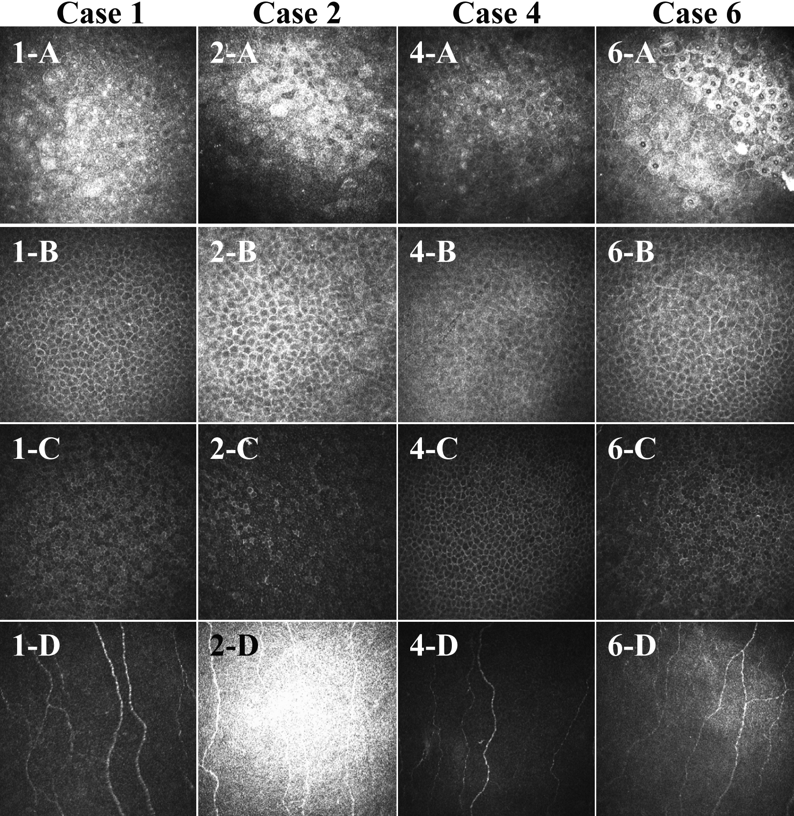
 Figure 3 of Chikama, Mol Vis 2008; 14:2333-2339.