Figure 2. In vivo confocal microscopic
images obtained with the HRTII-RCM system of all layers of the cornea
in a normal eye and the six cases of traumatic RCE. The images of the
first two columns represent areas of 400 µm×400 µm corresponding to the
superficial epithelial cell layer (A), wing cell layer (B),
basal cell layer (C), Bowman’s layer (D), shallow stroma (E),
mid stroma (F), deep stroma (G), and endothelium (H)
of the normal eye. Panel 1 (case 3) shows elongated and migrating
superficial cells. Panel 2 (case 2) shows gaps (arrows) in the
superficial epithelial layer due to epithelial defects. Panel 3 (case
3) shows the enlargement of basal epithelial cells (arrows). Panel 4
(case 2) shows the absence of subepithelial nerves as well as brightly
reflective granular structures at the bottom of the basal cell layer
and in Bowman’s layer. Panels 5a (case 4), 5b (case 6), and 5c (case 2)
show brightly reflective granular structures (arrows) in the wing cell
layer, in the basal cell layer, and in Bowman’s layer, respectively.
Panels 6a (case 2) and 6b (case 5) show activated keratocytes (arrows)
in the shallow stroma. Panel 7 (case 4) shows scattered fine particles
in the shallow stroma. Panel 8 (case 1) shows infiltrated cells, likely
neutrophils (arrows), in the mid stroma. Panel 9 (case 2) shows a small
number of keratoprecipitates (arrow) on the endothelium.
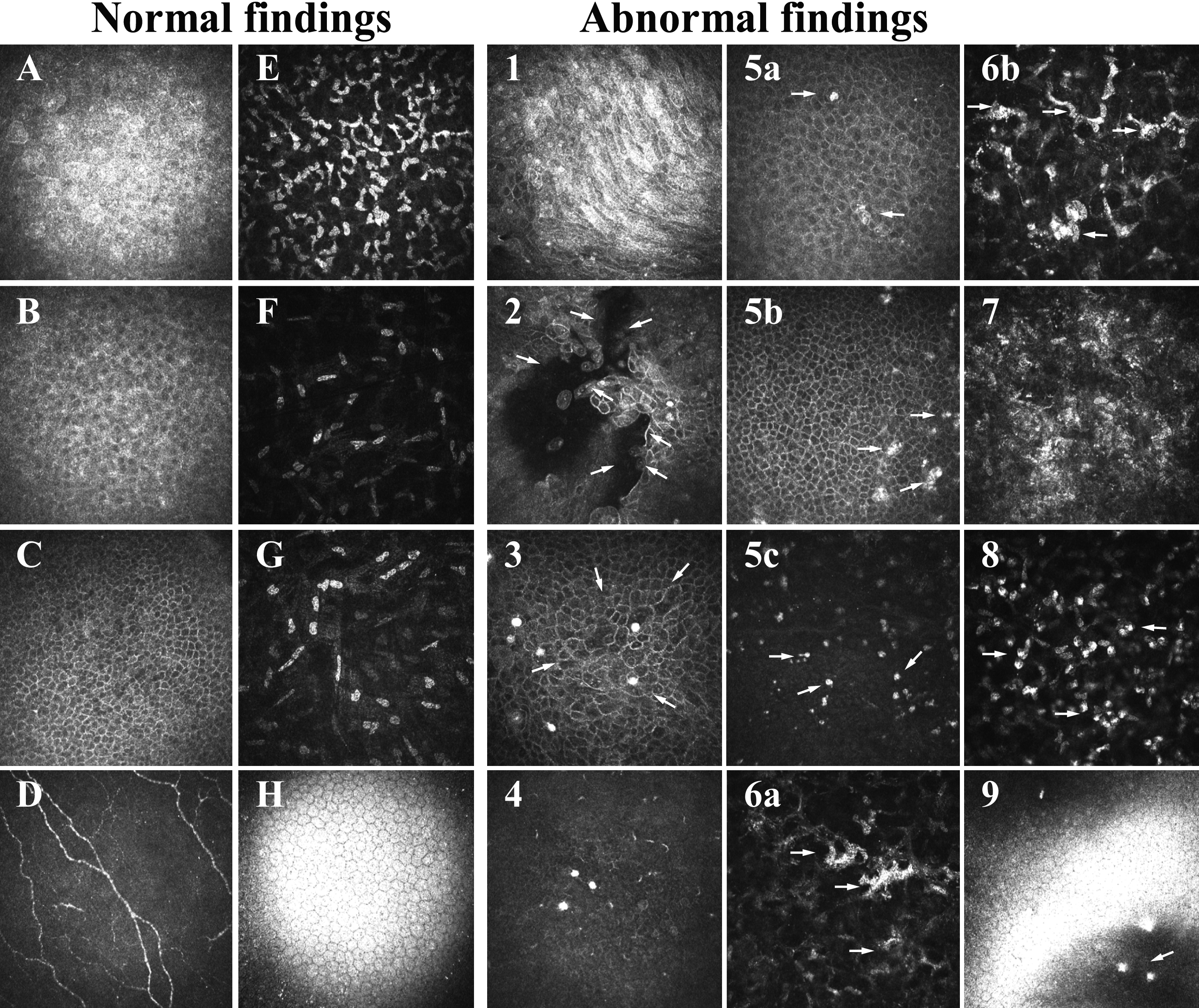
 Figure 2 of Chikama, Mol Vis 2008; 14:2333-2339.