Figure 1. Slit lamp biomicroscopic
findings of the six cases of traumatic RCE. The images of the first
column (A) were obtained with diffuse illumination. The images
of the second column (B) are higher power views obtained with a
combination of retroillumination and proximal illumination. The images
of the third column (C) show fluorescein staining.
Semitransparent or small white deposits were observed in all cases (B).
Corneal epithelial defects are stained green with fluorescein in cases
1 and 2 (C).
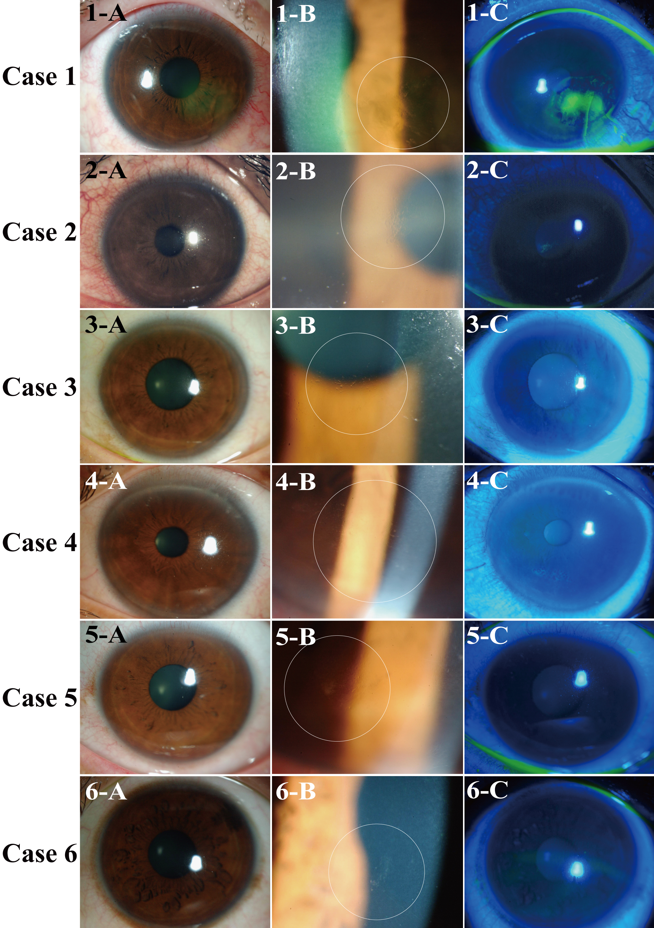
 Figure 1 of Chikama, Mol Vis 2008; 14:2333-2339.