Figure 1. Characteristic signs of XLRS. A: Fundus photograph of patient IV:2 of family 1 showing foveal schisis with spoke-wheel pattern. B: Fundus photograph of left eye of patient III:3 of family 3 showing bullous infero-temporal retinoschisis. C: OCT image (scan length: 6 mm, horizontal section) of patient IV:2 of family 1 showing foveal cystic retinoschisis in three
different retinal layers (marked by red arrows) and the color-coded foveal thickness maps showing the eccentric fixation.
GCL indicates ganglion cell layer, INL represents inner nuclear layer and PRL refers photoreceptor layer. D: OCT image of patient III:2 of family 5 showing foveal atrophy and the color-coded foveal thickness map showing the eccentric
fixation. E: Standard combined response of full-field ERG showing decreased b-wave amplitude (“negative type” ERG) related to normal (see
red arrow). F: Multifocal ERG with 61 first-order kernels showing decreased b-(P1) wave amplitudes in the central rings (marked by red).
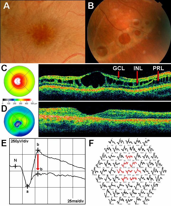
 Figure 1 of
Lesch, Mol Vis 2008; 14:2321-2332.
Figure 1 of
Lesch, Mol Vis 2008; 14:2321-2332.