Figure 4. Reprogrammed cultures lacked
vimentin+ cells that were Müller glia-like. A, B:
Immunostaining for vimentin (B) showed no cells that displayed
spindle-like processes (i.e., Müller glia-like) in the control culture
infected with Replication Competent Avian Splice (RCAS)-green
fluorescent protein (GFP), and A is a bright-field view of the
culture. C, D: Reprogrammed cultures (infected with RCAS-ash1)
also lacked vimentin+ cells with spindle-like processes (D);
C is a bright-field view of the culture. Arrows point to cells
with compact cell bodies discernible under Hoffman modulation optics. E-G:
A lack of vimentin+ cells that displayed spindle-like
processes in reprogrammed culture was not due to a lack of
reprogramming, because double-labeling showed that no vimentin+
cells spindle-like processes were detected (F) at places where a
large number of calretinin+ cells were present (G); E
is a bright-field view of the culture. Arrows point to calretinin+
cells. H and I are higher magnifications of F
and G, respectively. Scale bars represents 50 μm.
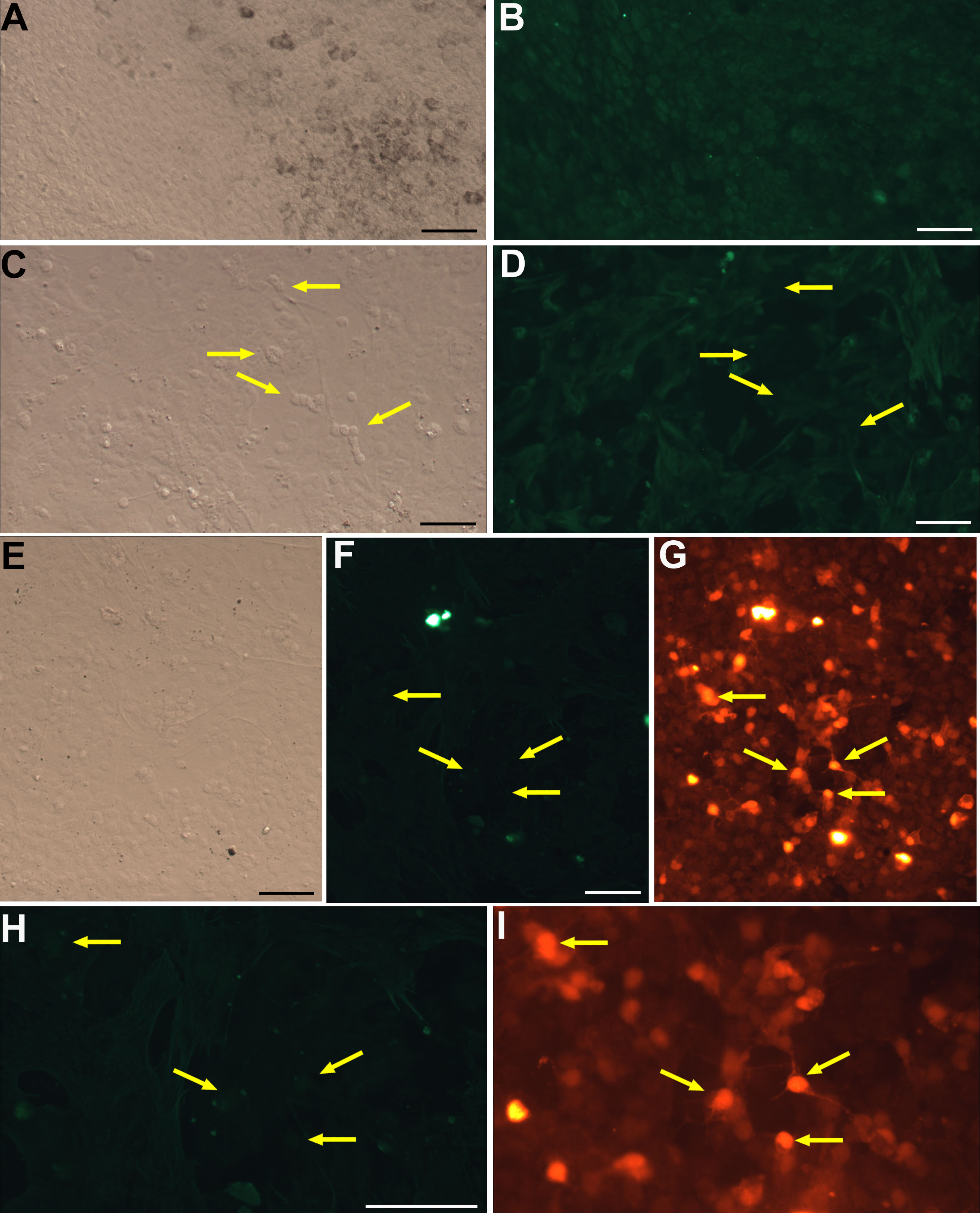
 Figure 4 of Mao, Mol Vis 2008; 14:2309-2320.