Figure 3. Neuron-like morphologies were
formed by reprogrammed cells in RPE cell cultures infected with RCAS-ash1
(or RCAS-ash1ΔCrb). A: Under bright-field, a
control retinal pigment epithelium (RPE) cell culture infected with
RCAS contained cells densely organized into a mono-layer. B:
After fluo-4 AM labeling, these cells were invisible with
epifluorescence, due to their low Ca2+ levels. C: A
calretinin+ cell in an RPE cell culture infected with
Replication Competent Avian Splice (RCAS)-achaete-scute homolog 1
(ash1) exhibited elaborate cellular processes, reminiscent of
neural processes. D: Reprogrammed cells in a RPE cell cultures
infected with RCAS-ash1 displayed neuron-like morphologies, as
revealed with fluo-4 AM labeling. E: Reprogrammed cells in RCAS-ash1ΔCrb-infected
culture also displayed neuron-like morphologies, as revealed with
transfection of with AAV-GFP DNA. Scale bars represents 50 μm.
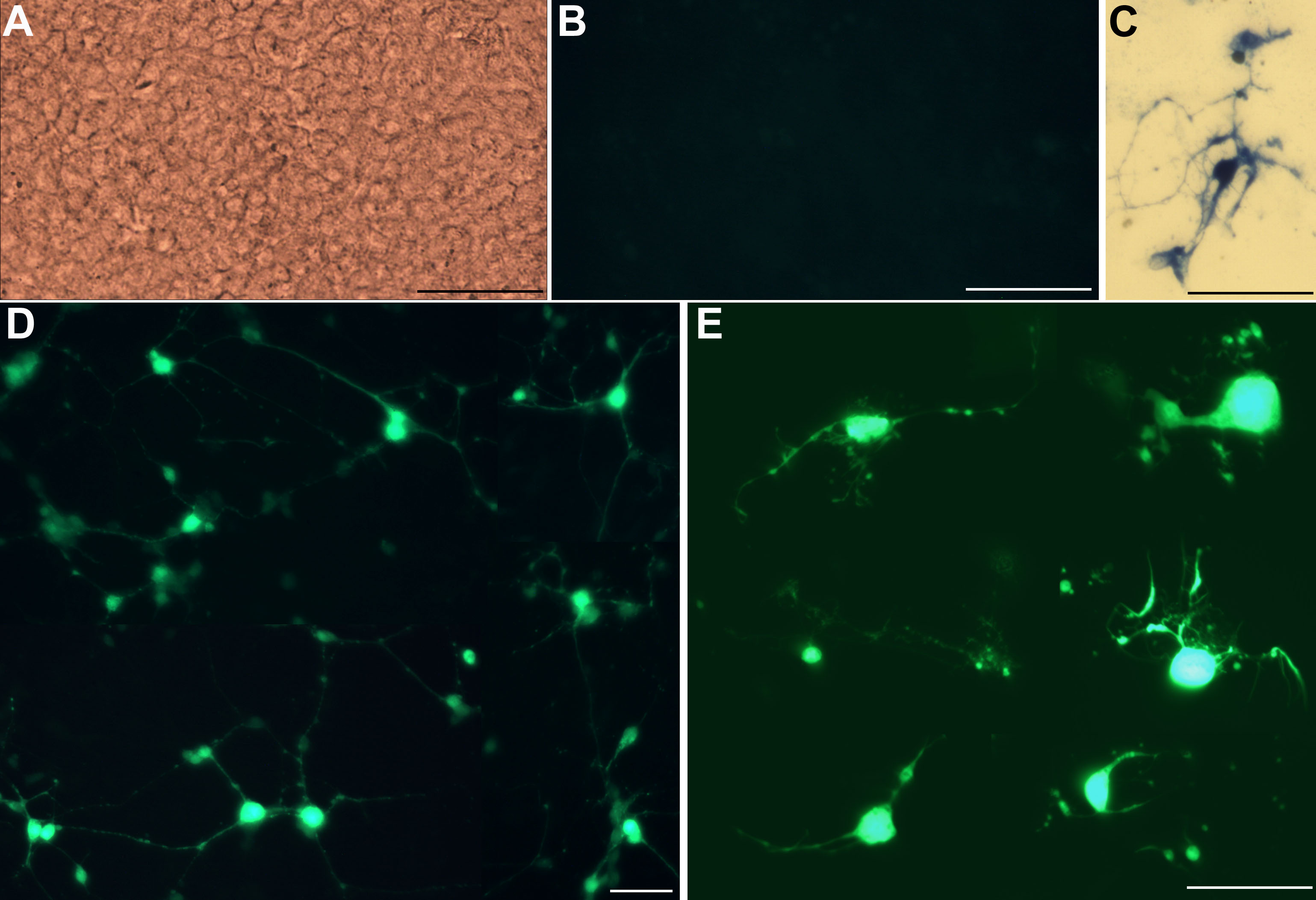
 Figure 3 of Mao, Mol Vis 2008; 14:2309-2320.