Figure 2. Neural markers were detected in
RPE cell cultures infected with RCAS-ash1 (or Replication
Competent Avian Splice (RCAS)-ash1ΔCrb) with
immunocytochemistry. A: Immunostaining for calretinin showed no
positive cells in a control culture infected by RCAS-green fluorescent
protein (GFP). B: A large of number of calretinin+ cells were
present in a culture infected by RCAS-ash1. C is a
higher magnification view of a section in B. Arrows point to
clusters of calretinin+ cells. D: MAP2+
cells were present in a culture infected by RCAS-ash1ΔCrb.
E: MAP2+ cells in RCAS-ash1ΔCrb-infected
culture displayed neuron-like morphologies. F-H:
Double-labeling for calretinin (F) and visinin (G) showed
that a small number of calretinin+ cells were also visinin+.
H is a merge of F and G. Arrowheads in F,
G, and H each point to a visinin+ cell that
lacked calretinin immunostaining. Scale bars represents 50 μm.
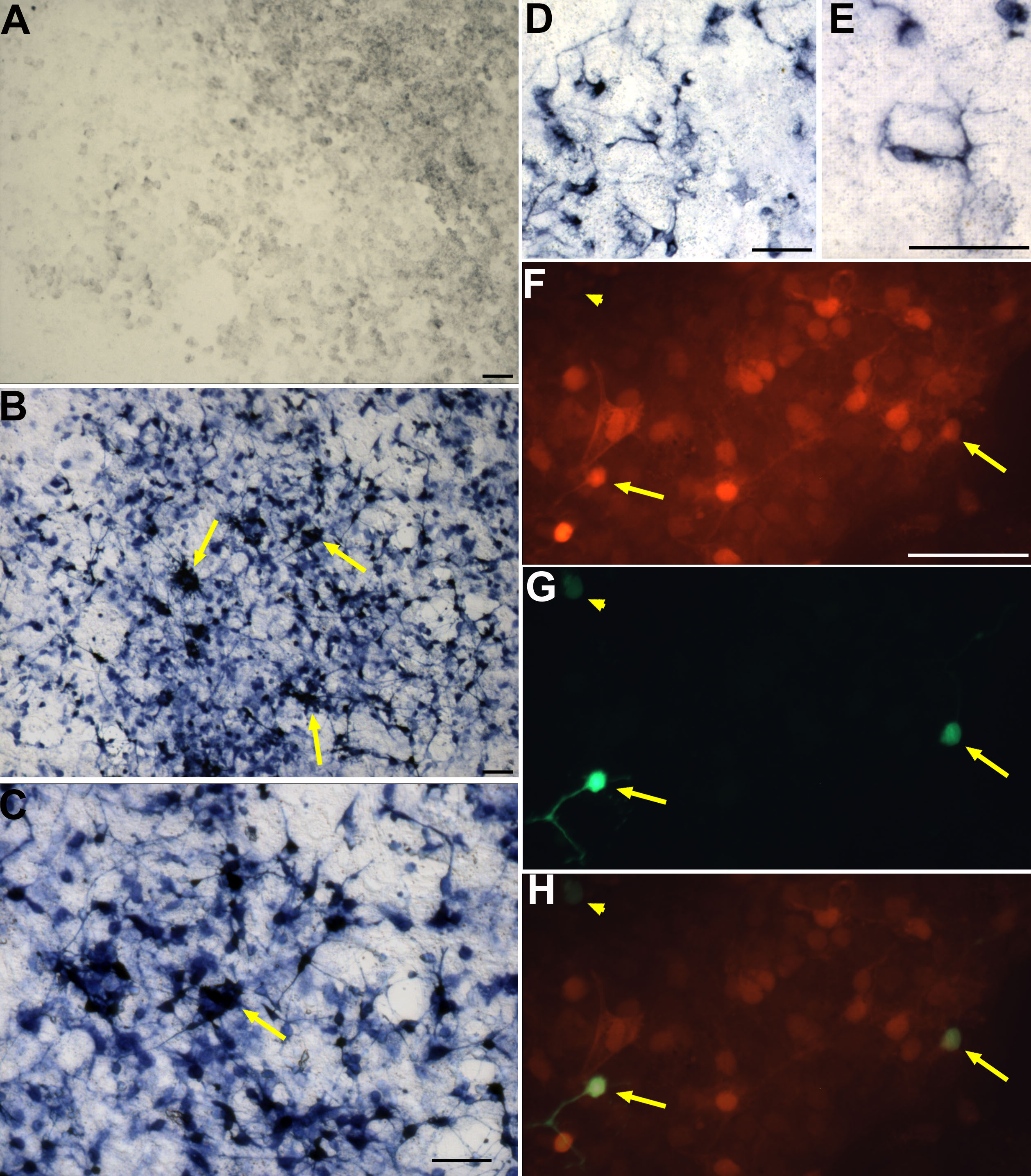
 Figure 2 of Mao, Mol Vis 2008; 14:2309-2320.