Figure 1. The appearance of RPE cell
culture infected with RCAS-ash1 differed from that of the
control. A: The control retinal pigment epithelium (RPE) cell
culture infected with Replication Competent Avian Splice (RCAS)-green
fluorescent protein (GFP; left) showed dark pigmentation, but the
experimental culture infected with RCAS-achaete-scute homolog 1 (ash1;
right) remained un-pigmented, except at a few places. B: The
control culture displayed a monolayer-appearance. C: The
experimental culture contained clusters or aggregates of cells. Arrows
point to cell clusters. Scale bars represents 100 μm.
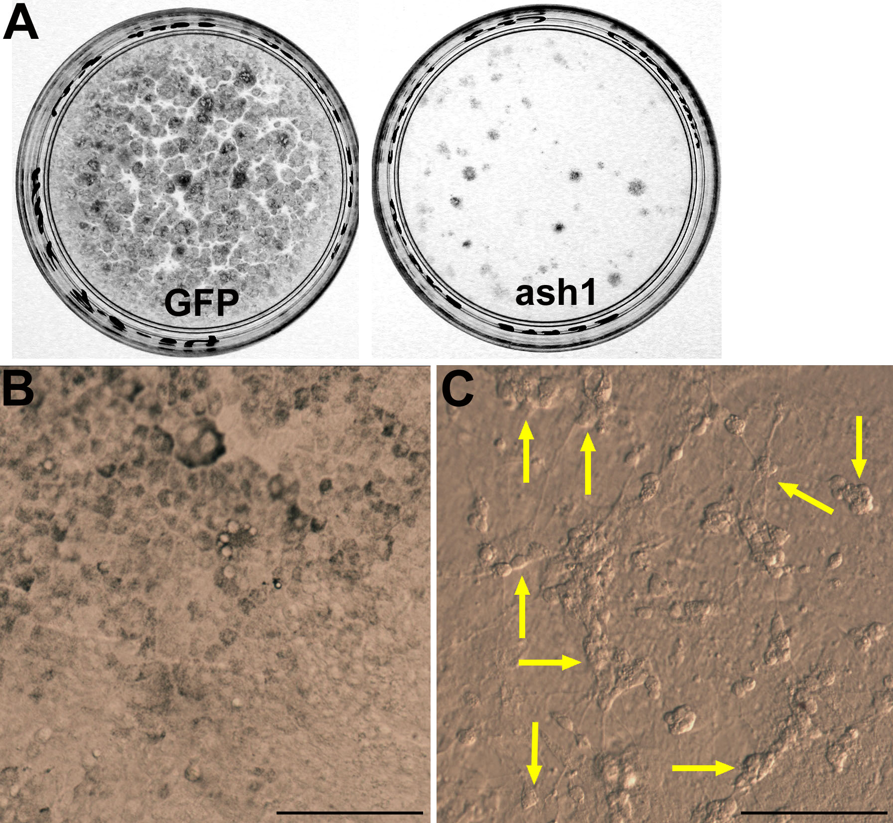
 Figure 1 of Mao, Mol Vis 2008; 14:2309-2320.