Figure 9. Expression pattern of a marker for endothelial-mesenchymal transition, α-smooth muscle actin, and that for fibrogenic reaction,
type I collagen, as well as PCNA in the healing, post-alklai burn, endothelium. The cells in the fibrous tissue posterior
to Descemet’s membrane in the control eyes were labeled for αSMA, the marker of myofibroblast or EnMT, as early as day 3 (A, arrows; compare to B). The cells in such a fibrous structure were markedly labeled at week 1 (C) and week 2 (E). Such labeled cells were not seen in the corneas of the Smad7-Ad group throughout the intervals of healing. Stromal cells
exhibited positive αSMA at week 2 (F). Type I collagen was stained as a marker of fibrous matrix accumulation. In the eyes of the control group, type I collagen
was detected in the matrix of fibrous tissue formed at week 1 (F) and week 2 (K) but not at day 3. No type I collagen immunoreactivity was observed posterior to the Descemet’s membrane in the eyes of the
Smad7-Ad group throughout the intervals (H,J,L). Nuclear staining of PCNA was more prominent in an eye of the Smad7-Ad group (N) than in an eye of the Cre-Ad group (M). Such a difference of PCNA staining was not seen between the groups at day 3 or at week 2 (data not shown). Bar, 50 μm.
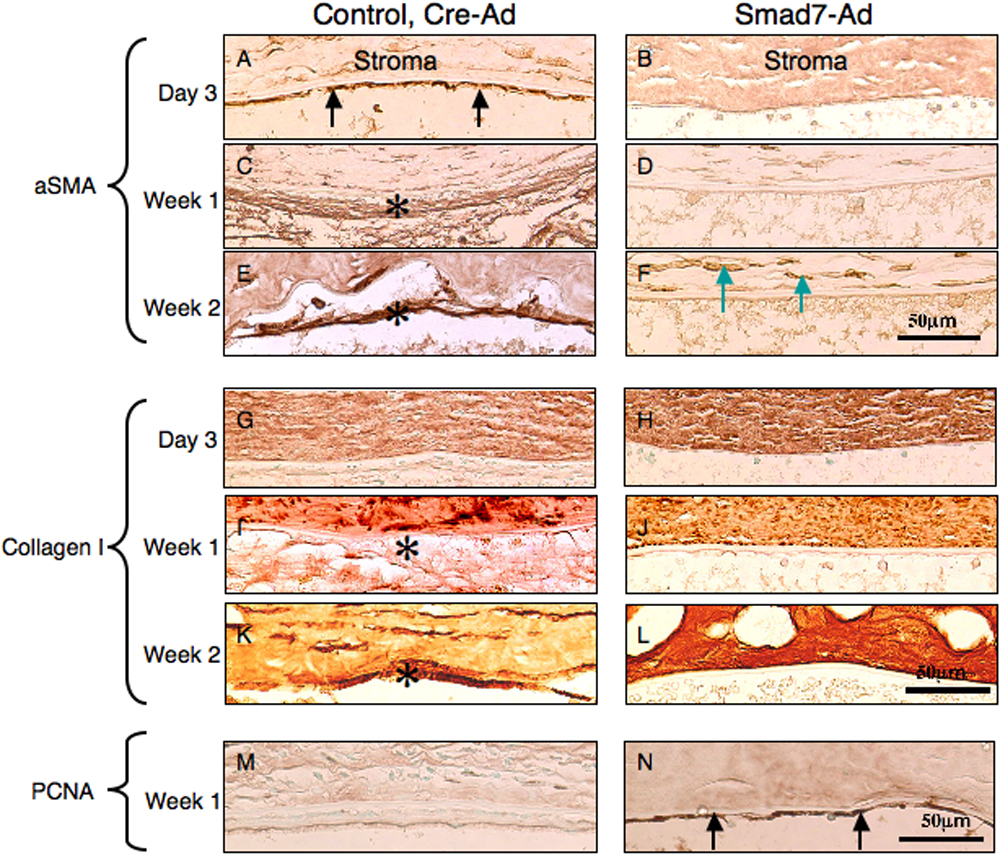
 Figure 9 of
Sumioka, Mol Vis 2008; 14:2272-2281.
Figure 9 of
Sumioka, Mol Vis 2008; 14:2272-2281.