Figure 8. Expression pattern of phospho-Smad2 in healing endothelium of an alkali-burned cornea. Immunohistochemistry showed that the
cells in the fibrous tissue in the control eyes were labeled for nuclear phospho-Smad2 at day 3 (A) and the number of such labeled cells increased in the fibrous tissue that formed posterior to Descemet’s membrane at week
1 (C) and week 2 (E). Phopsho-Smad2 positive cells were not seen in the endothelial layer of Smad7-Ad eyes at each time point (B,D,F). The finding indicates that exogenous Smad7 might block Smad signaling. Bar, 50 μm.
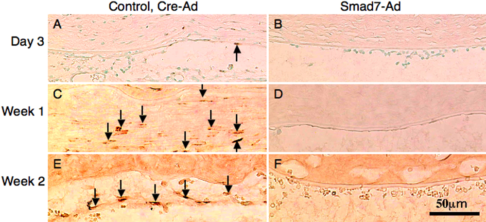
 Figure 8 of
Sumioka, Mol Vis 2008; 14:2272-2281.
Figure 8 of
Sumioka, Mol Vis 2008; 14:2272-2281.