Figure 7. Hematoxylin and eosin staining of the corneal endothelium during healing after alkali exposure. Histology showed that there
was abnormal accumulation of fibrous tissue (asterisk) posterior to the Descemet’s membrane at day 3 (A), week 1 (C), and week 2 (E) in the eyes of the control Cre-Ad group while such findings were not observed in the eyes of the Smad7-Ad group at each
time point (B,D,F). Inflammation in the stroma was also less severe in a Smad7-Ad treated cornea compared to a control cornea at day 3. Bar,
50 μm.
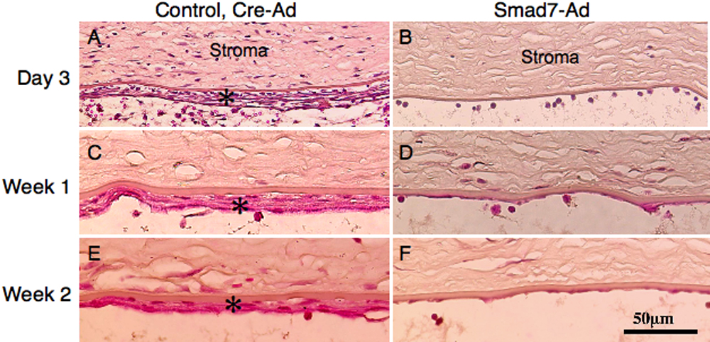
 Figure 7 of
Sumioka, Mol Vis 2008; 14:2272-2281.
Figure 7 of
Sumioka, Mol Vis 2008; 14:2272-2281.