Figure 11. Expression and subcellular
localization of claudin 20 in culture. RPE was isolated from E7 or E14
embryos and cultured with SF2 in the basal medium chamber and either
SF3 or rcSF3 in the apical medium chamber. The cultures were labeled
for claudin 20 (A) or double labeled for claudin 20 and ZO-1 (B).
Under standard optics (A), claudin 20 was observed along the
lateral membranes in each culture. When E7 RPE was cultured with SF3 in
the apical medium chamber, a vesicular distribution was less evident
when the RPE was cultured with rcSF3 in the apical medium chamber.
Confocal microscopy (B) revealed the distribution of claudin 20
and ZO-1 in the XZ plane. Claudin 20 colocalized with ZO-1 in a
junctional complex at the apical end of the lateral membranes, as
indicated by the yellow in the merged image. In (B), arrowheads
that point up indicate basal membrane, while arrowheads that point down
mark apical membrane. The scale bar represents 10 μm.
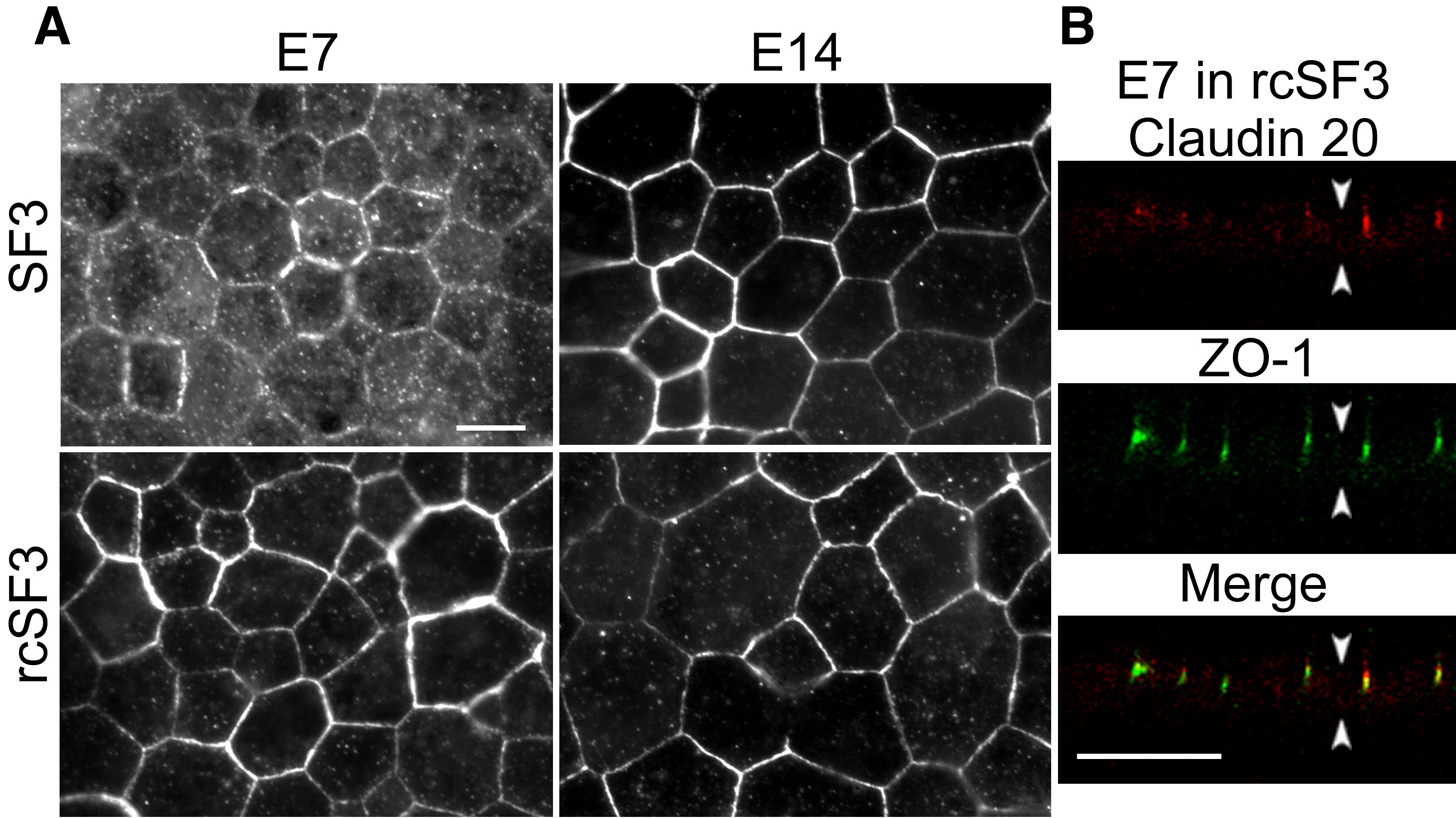
 Figure 11 of Sun, Mol Vis 2008; 14:2237-2262.