Figure 10. Expression and subcellular
localization of claudin 20 and ZO-3 in vivo. Sheets of RPE and choroid
were isolated from E10 and E18 eyes and fixed. The distribution of
claudin 20 or ZO-3 was revealed by indirect immunofluorescence, as
described in Methods. Neither protein could be detected in E10 RPE, but
a clear signal was observed along the lateral membranes in E18 RPE. No
signal was detected when the relevant peptide antigen was used to
compete for binding of the antibody (data not shown). The scale bar
represents 10 μm.
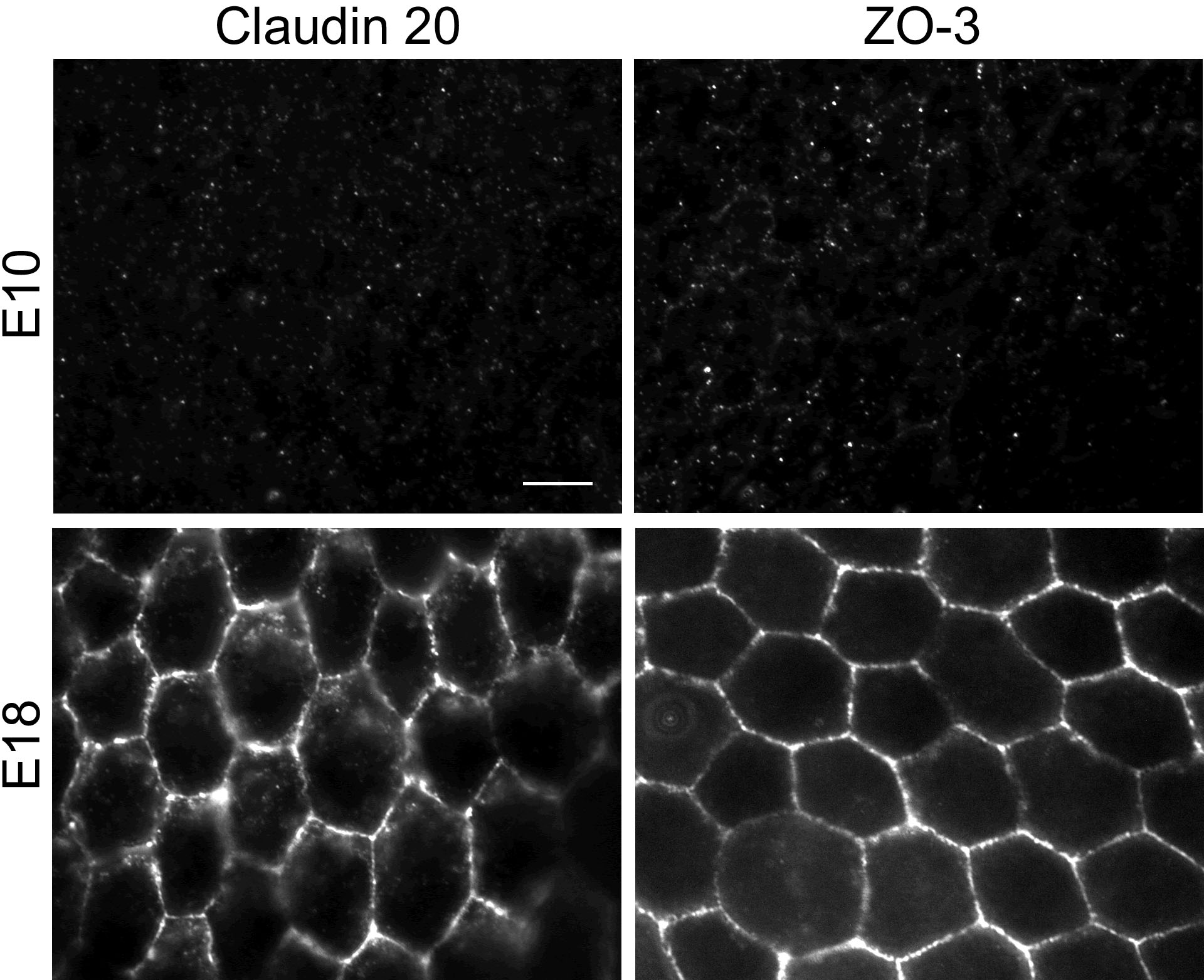
 Figure 10 of Sun, Mol Vis 2008; 14:2237-2262.