Figure 5. Conjunctival impression cytology stained by cresyl violet at D1. PBS (A) presented normal aspects of the conjunctival epithelium with no inflammatory infiltration. BAK Sol (B) induced numerous polymorphonuclear inflammatory cells with almost no normally shaped epithelial cell visible. BAK Em (C) and CKC Sol (D) both showed epithelial damage with inflammatory infiltration. CKC Em-instilled (E) rabbit eyes presented normal epithelial patterns without inflammatory infiltration. (original size 40×).
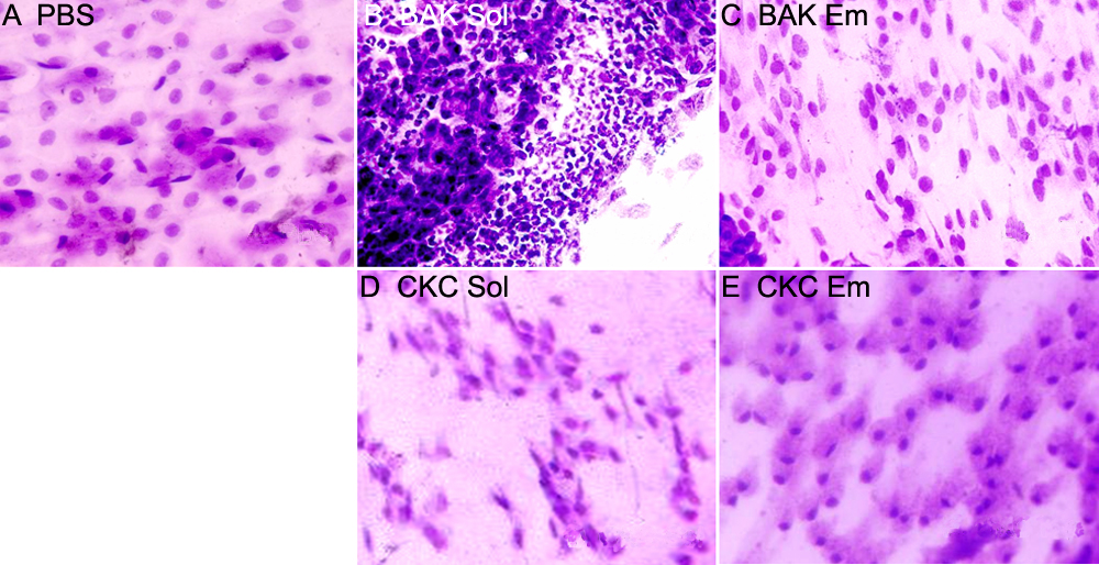
 Figure 5 of
Liang, Mol Vis 2008; 14:204-216.
Figure 5 of
Liang, Mol Vis 2008; 14:204-216.