Figure 3. HRT II IVCM images of rabbit ocular surface. HRT II IVCM images of rabbit ocular surface after PBS (A), BAK Sol (B), BAK Em (C), CKC Sol (D), and CKC Em (E) instillations at D1 are displayed. Results are shown in the superficial epithelium (line 1), basal epithelium (line 2: 10–15
μm from the superficial epithelium layer), anterior stroma (line 3: 50–65 μm from the superficial epithelium layer), and conjunctival
substantia propria (line 4: 60–90 μm from the superficial epithelium layer). BAK Sol-receiving eyes showed the greatest damage
in the epithelium and the greatest inflammatory infiltration in the basal epithelium and anterior corneal stroma. BAK Em and
CKC Sol induced intermediate toxicity. These three groups induced inflammatory cells rolling in conjunctival blood vessels.
CKC Em presented almost the same aspects in all ocular surface structures as the PBS-instilled group. The scale bar indicates
100 μm.
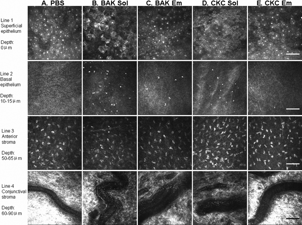
 Figure 3 of
Liang, Mol Vis 2008; 14:204-216.
Figure 3 of
Liang, Mol Vis 2008; 14:204-216.