Figure 8. Expression and death assessment
in the retina following subretinal injection and electroporation of
pVAX-tdTomato. A: Cross-sections of the retina were cut with a
cryostat. Red fluorescence was detected in a single cell found in the
RPE layer. This image is the red channel from a confocal image. B:
TUNEL staining (green channel) was done in the same section. This panel
shows three TUNEL-positive nuclei in a field of roughly 400 nuclei in
the PhR cell layer in the image. C: A and B
were merged and show the DAPI channel (blue) of the same section. The
scale bar represents 50 μm.
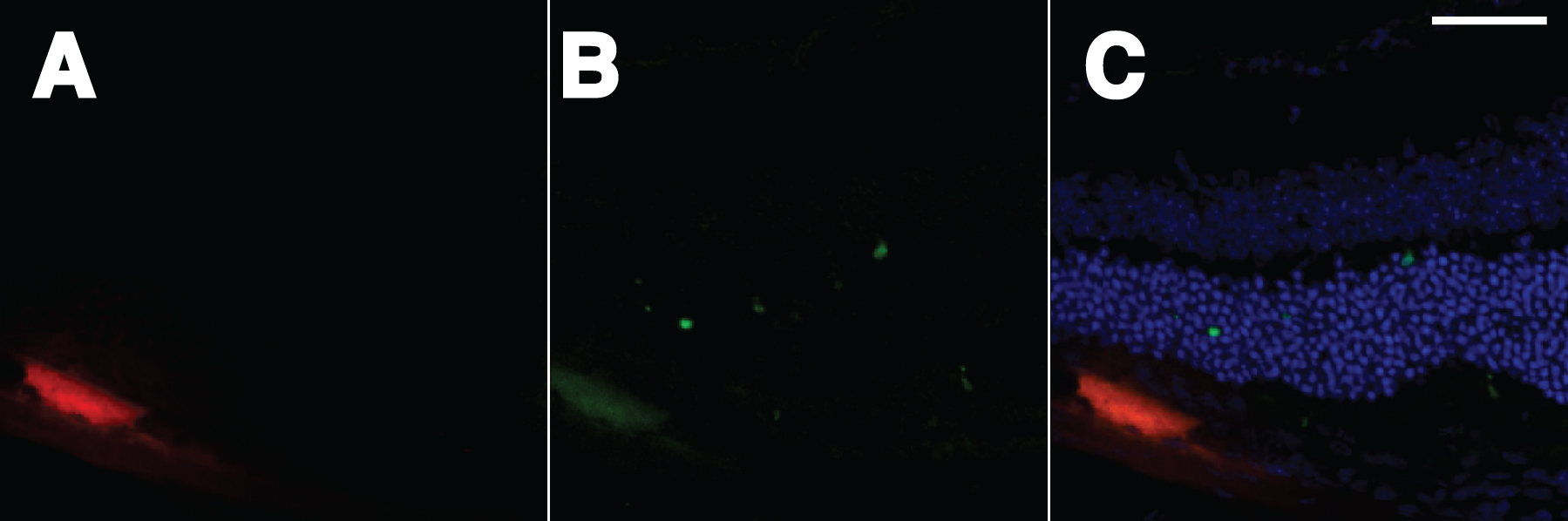
 Figure 8 of Johnson, Mol Vis 2008; 14:2211-2226.