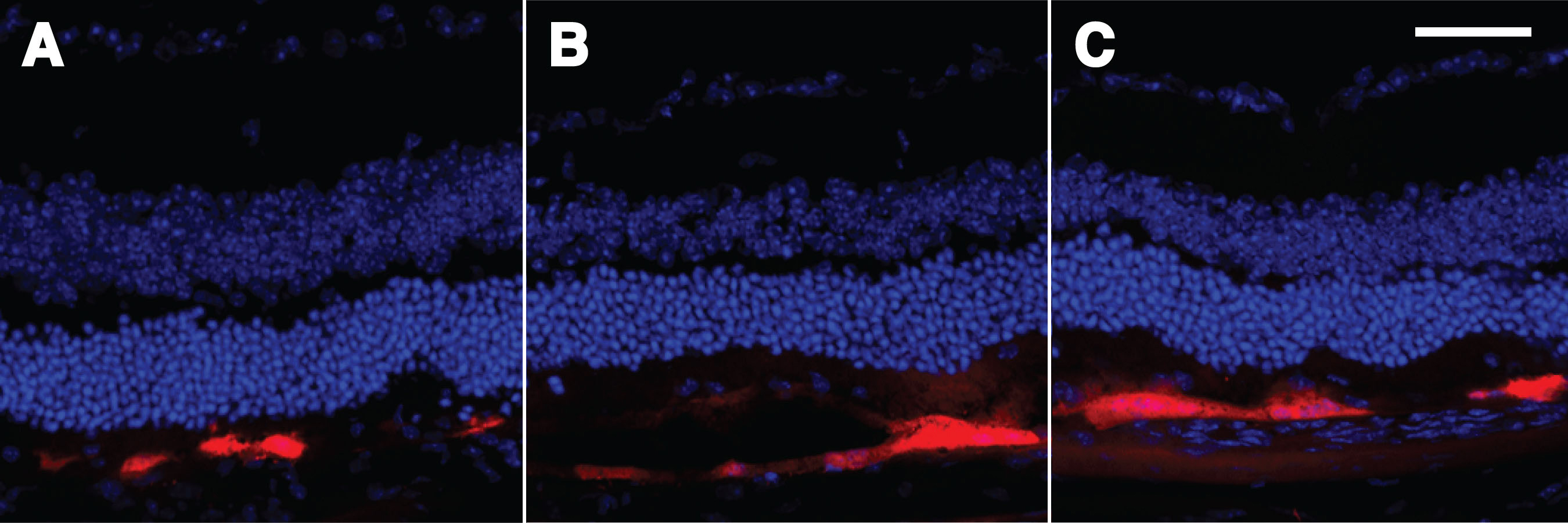
Figure 7. Electroporation of the mouse eye
viewed in cross section.
A,
B, and
C:
Demonstrated are the results of representative experiments with
optimized electroporation conditions after the subretinal injection of
pVAX-tdTomato [
52].
The cross-sections were cut with a cryostat and nuclei stained with
DAPI.
A,
B, and
C all illustrate specific
expression of tdTomato in the RPE cell layer. The red RPE cells each
appear to subtend a region of about 50 nuclei in the outer nuclear
layer, consistent with the known relationship of each RPE cell
supporting numerous PhR cells. The scale bar represents 50 μm.
 Figure 7 of Johnson, Mol Vis 2008; 14:2211-2226.