Figure 6. Expression of a reporter gene in
RPE cells; subretinal injection and electroporation under optimized
conditions. Red fluorescence of tdTomato expression was observed
following subretinal injection and electroporation under optimized
conditions; individual RPE cells were resolved. The cells include
neighbors with and without tdTomato fluorescence (red). About 30% of
the cells in electroporated area were positive for tdTomato gene
expression. This field represents the RPE cells located directly over
the anode. TdTomato-expressing cells exhibited polygonal shapes
characteristic of normal RPE cells. The electroporation conditions
employed were 50 V, 2 mm gap between electrodes, 1 ms pulse duration,
and 10 pulses at 1 s intervals. A: In this schematic of the
eye, the bleb is in green and the positive electrode in red. The black
electrode represents the negative electrode. B: Presented is a
wholemount of the eye after bleb formation but without voltage applied
to the electrodes. The edges of the flatmounts are outlined in white
and form a floret shape. The center of the floret corresponds to the
retina while the outer half of the “petals” correspond to the cornea. C:
Shown is a wholemount of the eye following subretinal injection and
electroporation under the optimized condition. A focused patch of red
fluorescent cells is evident near the center of the floret. Each dot
represents a separate RPE cell. This region of the retina corresponds
to the bleb and the location of the anode. D: High
magnification of the fluorescent region shows about 30% of the RPE
cells manifesting tdTomato fluorescence. E: Close-up of a
cluster of tdTomato fluorescence in cells reveals a cobblestone or
polygonal shape. F: Shown is a close-up of a single binucleate
RPE cell. In B and C, images are about 9 mm across. The
scale bar in D represents 50 μ. The scale bars in E and
F represent 25 μm.
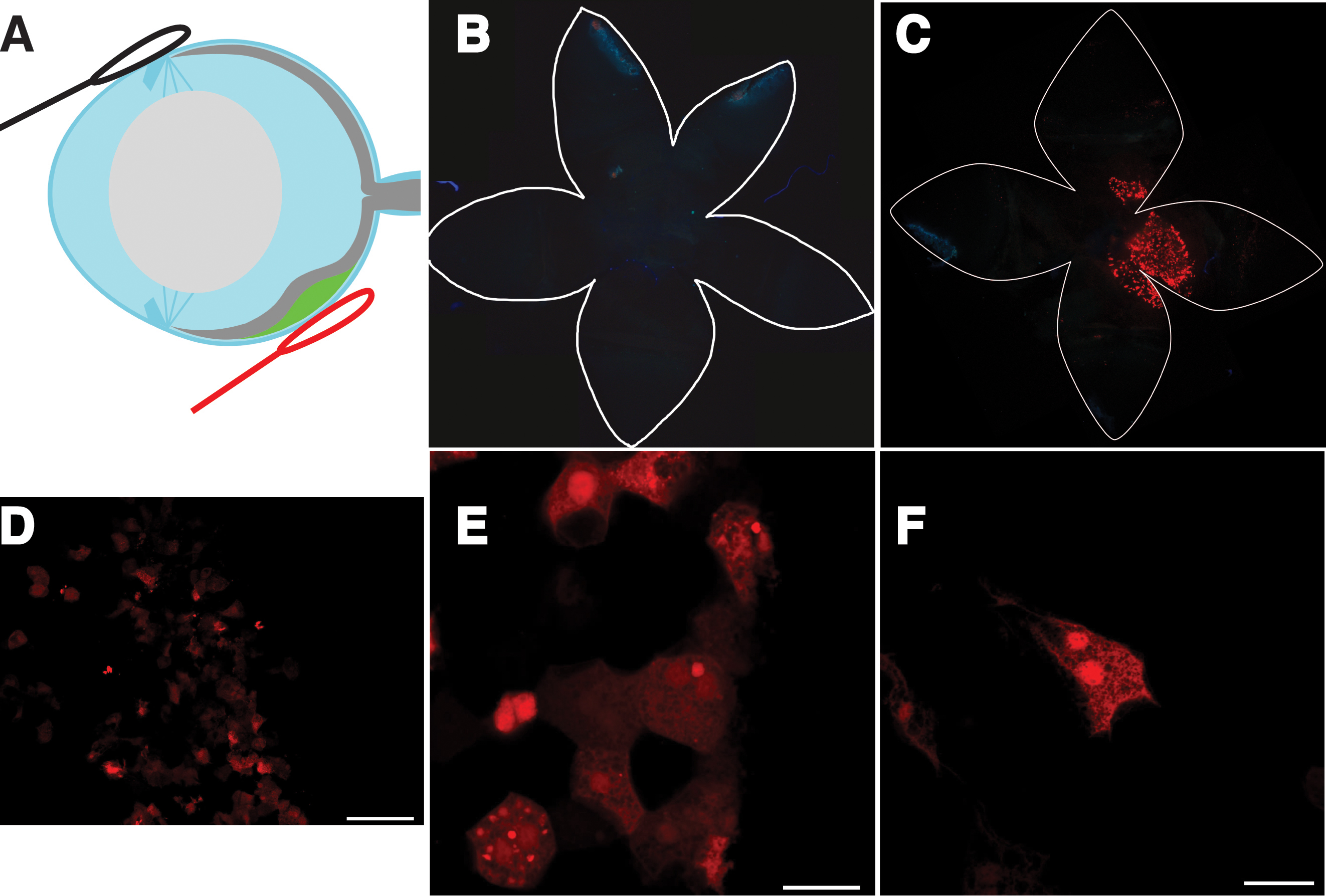
 Figure 6 of Johnson, Mol Vis 2008; 14:2211-2226.