Figure 4. Schematic diagram of PITX2a and amino acid sequence alignments of PITX2 homeodomain with other homeodomain proteins. A: The structure of the PITX2a isoform. The black region represents the homeodomain (HD). B: Alignments of the human PITX2 homeodomain and related homeodomain-containing proteins (the three α-helices are also indicated)
are shown. The arrows indicate the previously characterized mutations within the helix 3 of PITX2a homeodomain. The tryptophan
residue at position 86 is conserved among these homeodomain proteins.
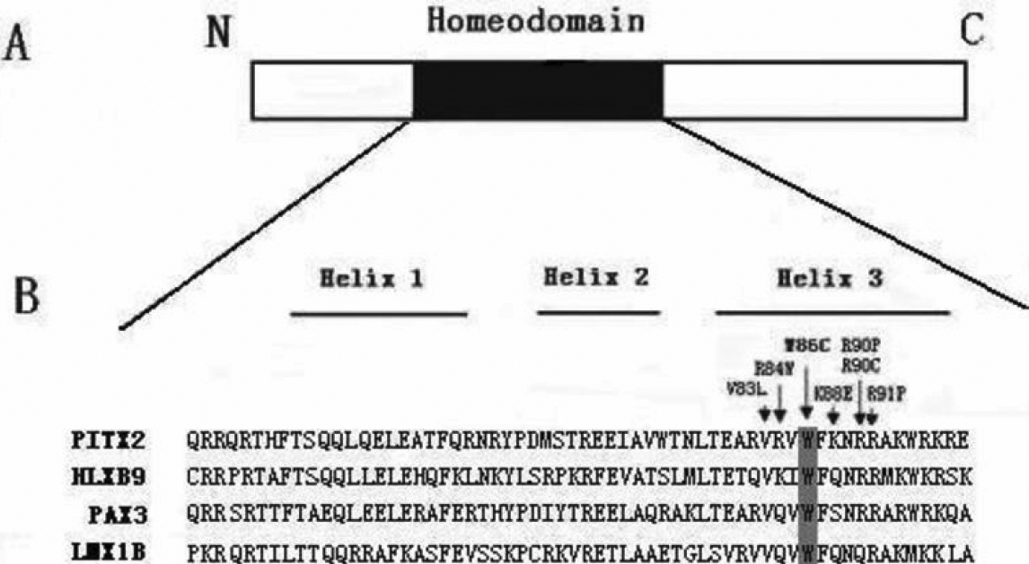
 Figure 4 of
Li, Mol Vis 2008; 14:2205-2210.
Figure 4 of
Li, Mol Vis 2008; 14:2205-2210.