Figure 4. Phase appearance and marker
protein expression of cultivated corneal limbal epithelial cells,
keratocytes, and endothelial cells. The rabbit corneal limbal
epithelial cells (A), keratocytes (B), and endothelial
cells (C) were cultivated for one week to confluence on culture
dishes, primary (10X). The protein of keratin 3 was specially expressed
in the cultivated corneal limbal epithelial cells while vimentin
expression was highest in the keratocytes. Aquaporin A was only
detected in the endothelium (D).
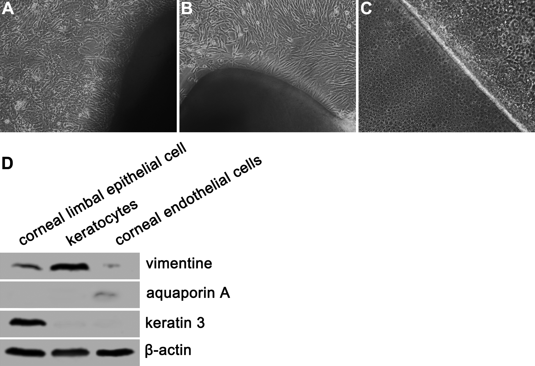
 Figure 4 of Xu, Mol Vis 2008; 14:2180-2189.