Figure 2. Histological characteristics of the ACMP. HE stained histological sections showed that the major immunogenic porcine corneal
epithelial cells, keratocytes, and endothelial cells were completely removed in (A; 10X) and (B; 40X). The epithelial cells, keratocytes, and endothelial cells would be detected on each layer of a normal porcine cornea
(C; 10X) and (D; 40X). SEM images of the ACMP showed that the collagen fibers and fibers inter-connecting to network had formed collagen
bundles, which were regular and parallel to the corneal surface (E; 5,000X) and (F; 20,000X). These were similar to the normal porcine cornea matrix.
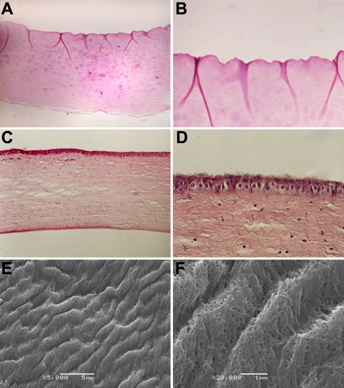
 Figure 2 of
Xu, Mol Vis 2008; 14:2180-2189.
Figure 2 of
Xu, Mol Vis 2008; 14:2180-2189.