Figure 6. Apoptosis assay: optic nerve,
after crush and rAION. A: Normal optic nerve; no apoptotic
cells are detected. B: Positive control. C:
TUNEL-positive (red) cells along the optic nerve 3 days following crush
injury of wild type mice. D: TUNEL staining of optic nerve of
GFP-CNPase mice 3 days following crush showing positive staining.
Oligodendrocytes apoptotic cells are yellow (green for GFP and red
represent positive staining for apoptosis), suggesting that
oligodendrocytes in the optic nerve proximal to the globe undergo
apoptosis 3 days following induction of crush injury. E: The
whole optic nerve of same GFP-CNPase mice (D) is shown. F:
Optic nerve head, 3 days following crush injury; showing hemorrhage
(asterisk), immediately posterior to the globe. G: Same damaged
area at higher magnification, demonstrating loss of oligodendrocytes
and focal hemorrhagic area. H: Optic nerve head 3 days
following rAION induction, showing preserved architecture of retina and
intraocular optic nerve, without apoptotic cells. I: Same nerve
as H, 3 days after rAION, demonstrating the anterior segment of
the optic nerve behind the globe. Note few TUNEL-positive cells (red
staining, arrows) at the center of the anterior optic nerve.
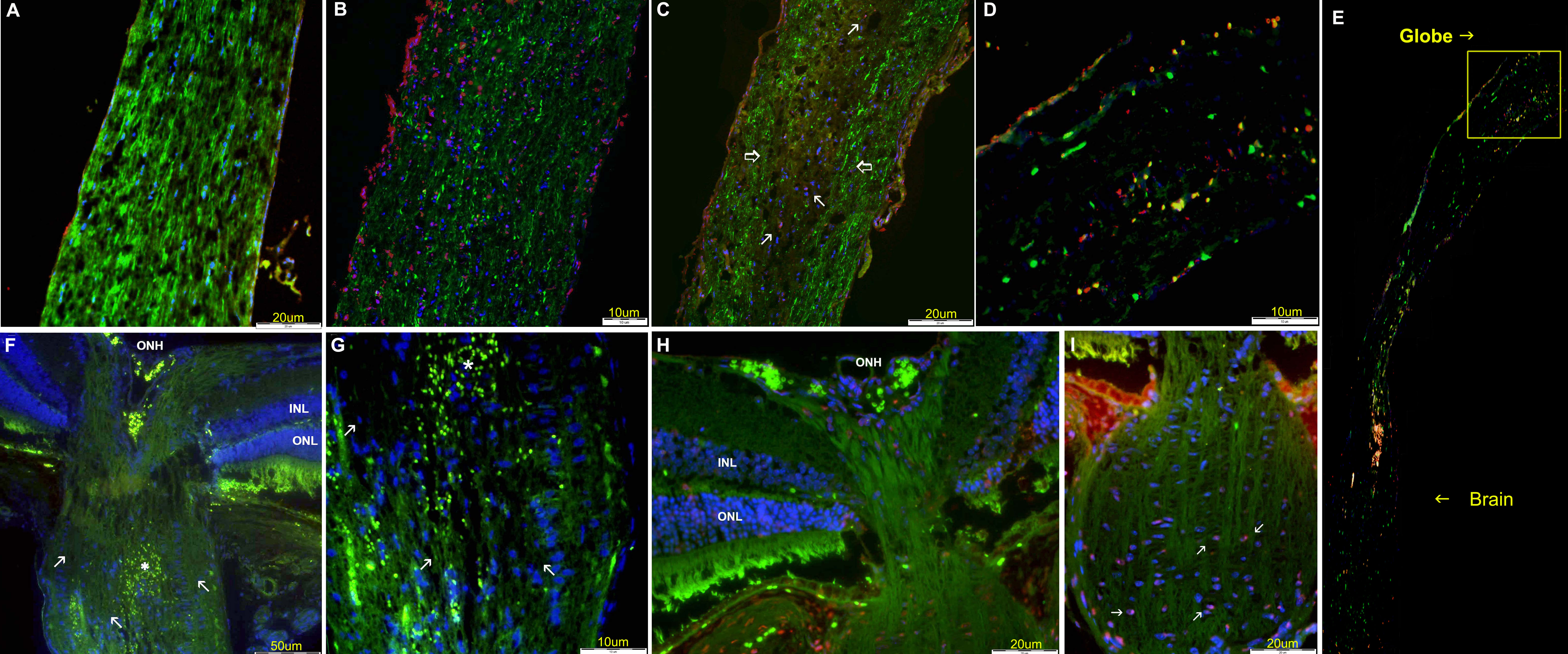
 Figure 6 of Dratviman-Storobinsky, Mol Vis 2008; 14:2171-2179.