Figure 2. RGC loss in flat-mounted retinas
of Thy1-CFP mice at various intervals after crush injury. A:
Control retina; no damage is detected. B: Four days after crush
injury; approximately 20%–30% RGC cell loss was detected. C:
Seven days after crush injury; 50% RGC loss was detected. D:
Fourteen days after crush injury; maximal 75% RGC loss can be detected.
Note the diffuse loss of the labeled cells.
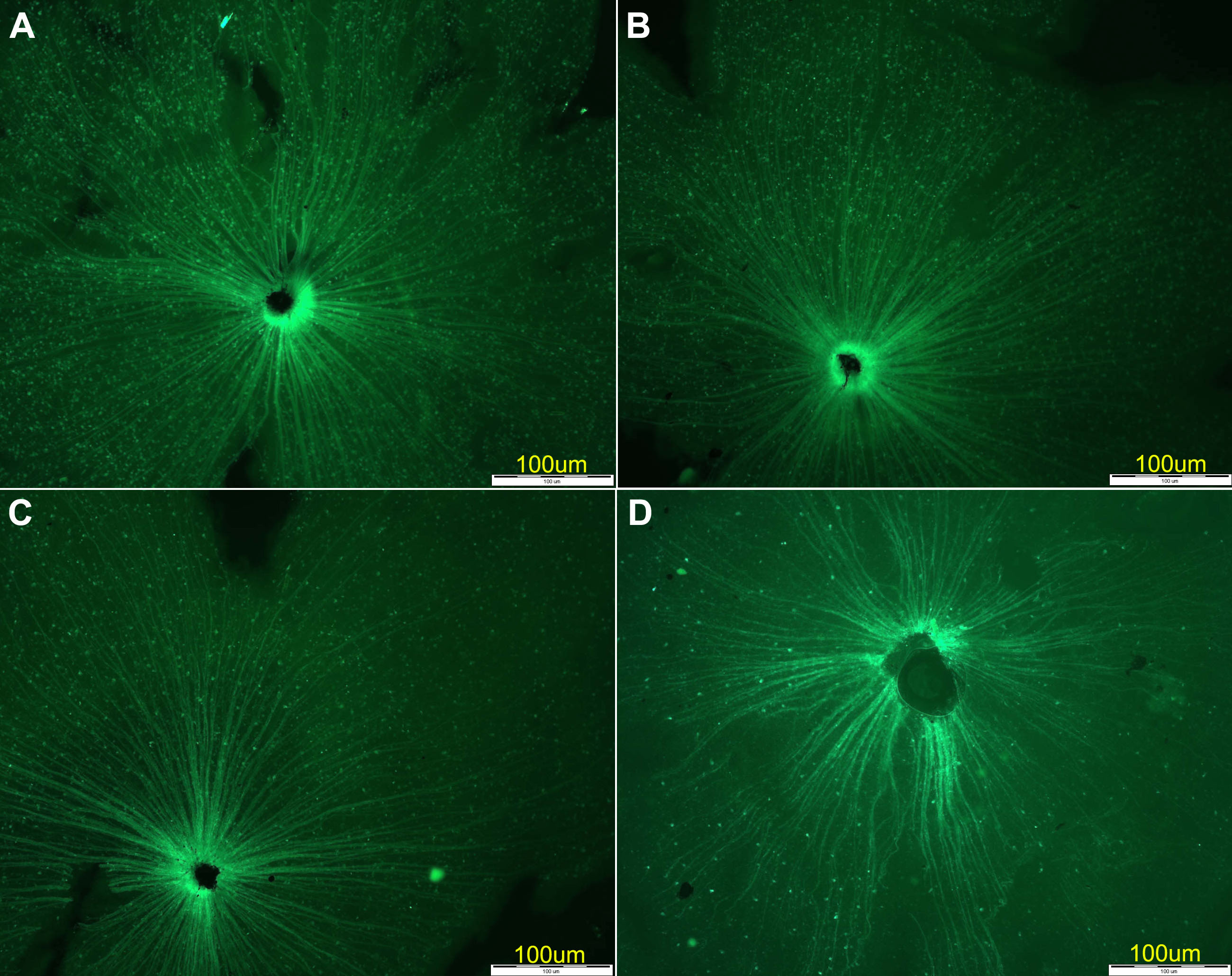
 Figure 2 of Dratviman-Storobinsky, Mol Vis 2008; 14:2171-2179.