Figure 8. Cardiac M-mode images of the
heart in 24, 48, 72, and 120 hours post fertilization embryos. The
bright signal was created by blood within the heart. Note the increase
in heart rate with development, as well as the development of two
chambers at 72 hours post fertilization (hpf). The heart rates observed
in the m-mode images are 47 beats per min (bpm) in the 24 hpf embryo,
157 bpm in the 48 hpf embryo, 219 bpm in the 72 hpf embryo, and 250 bpm
in the 120 hpf embryo.
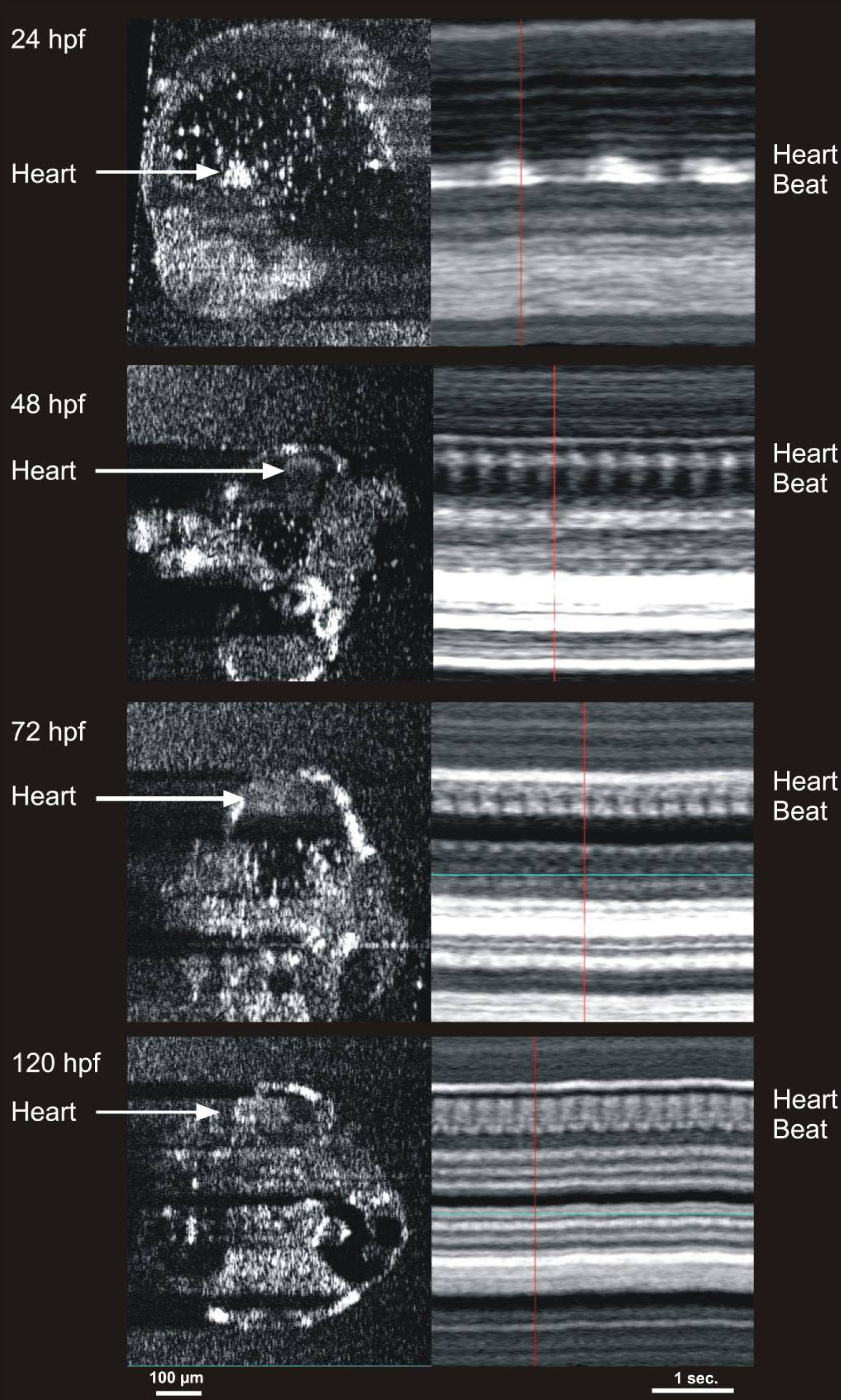
 Figure 8 of Kagemann, Mol Vis 2008; 14:2157-2170.