Figure 5. Stereo pairs of images reveal
internal structures in whole embryos in three dimensions. These panels
provide visualization of three dimensional (3D) data in a printed
image. Such data are best visualized interactively, rotating the
projected data manually to obtain an optimal view of the structure of
interest; impossible in printed image (see also Figure 2). Panel A
contains a 3D crossed-stereo image of a 24 hpf embryo. To view, gently
cross your eyes until 3 images appear, and focus on the image in the
middle. The image on the far right of panel A is the same
image, and can be viewed with red/blue 3D glasses to visualize the
embryo in 3D. Panel B contains a crossed-stereo pair and
red/blue stereo image of a 24 hpf embryo. Panel C contains a 72
hpf stereo-pair and red/blue 3D image. In panels A-C,
the embryo appears to be facing out of the image. Panel D
contains a stereo-pair and red/blue 3D image of a 120 hpf embryo facing
into the image.
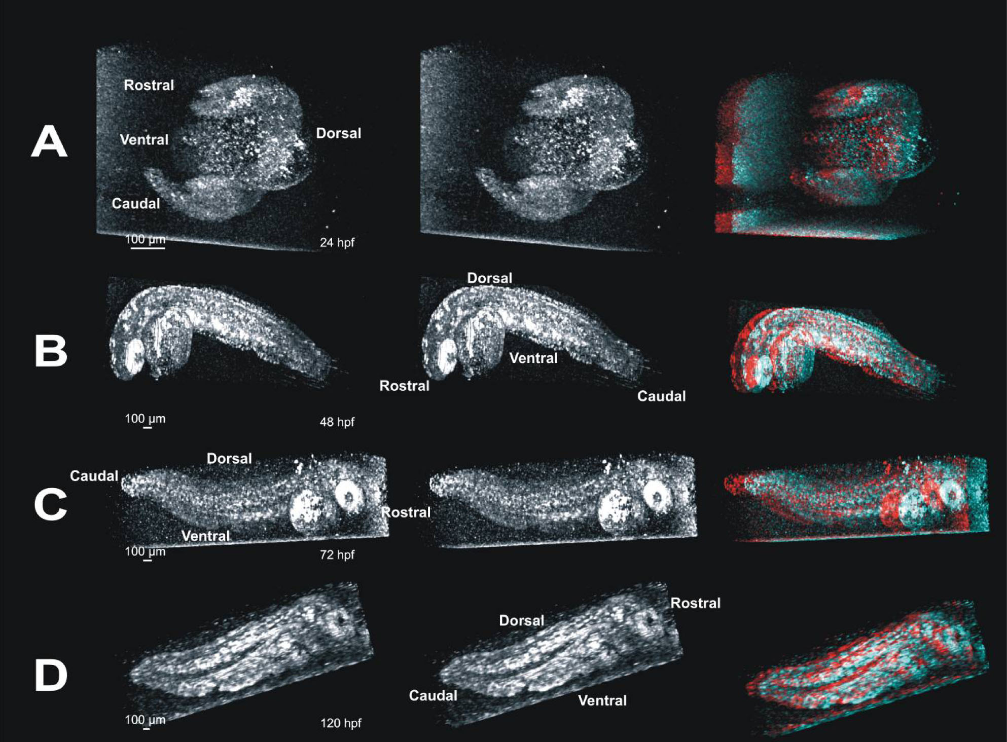
 Figure 5 of Kagemann, Mol Vis 2008; 14:2157-2170.