Figure 2. Rotation of a 3D image dataset
of a 120 hpf embryo. SD-OCT can acquire a 3 dimensional quantitative
description of the tissues within zebrafish embryos. These data allow
the non-invasive visualization of the entire animal, as well as
cross-sectional slices in any orientation through the animal at any
stage of development, all without sacrificing the animal or noticeably
impeding development. This gross anatomical visualization of the
zebrafish was rendered in 3D-View, using the maximum intensity
projection display.
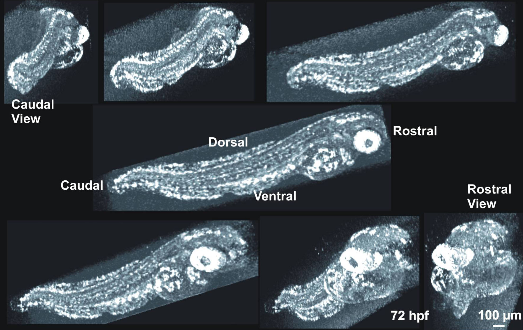
 Figure 2 of Kagemann, Mol Vis 2008; 14:2157-2170.