Figure 1. The appearance of the 72 hpf
zebrafish with millimeter ruler, at magnification, and as observed by
SD-OCT provided for appreciation of its small size. Zebrafish embryos
were embedded in 1% agarose gel in an inverted microscopy Petri dish (A,
B; B shows a magnified region of A). Embryos were scanned
in three dimensions (3D), and reflectance of internal structures
quantified. C-mode sections of the 3D data set could be isolated and
tissue reflectance within a slice is displayed (C).
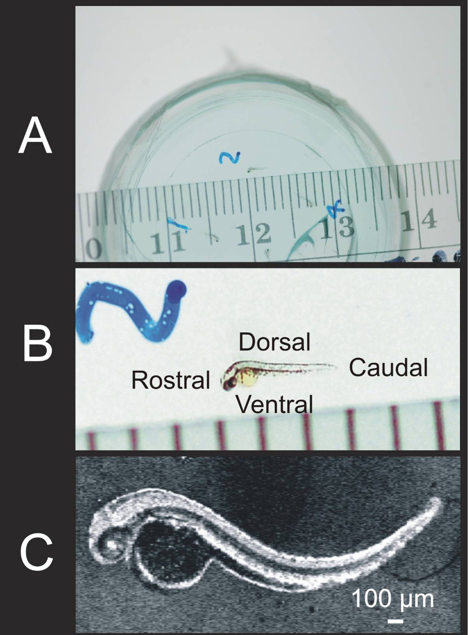
 Figure 1 of Kagemann, Mol Vis 2008; 14:2157-2170.