Figure 6. Immunofluorescence staining of cryosections from 6 day cocultured explants. Antibodies against glial fibrillary acidic protein
(GFAP; red) and cellular retinaldehyde-binding protein (CRALBP; green) were used. Confocal images revealed an increased expression
of GFAP and a decreased CRALBP expression in the neuroretinal external layers. Müller cell processes crossed the outer limiting
membrane (OLM; arrows). The cell bodies of GFAP+ astrocytes remained in the nerve fiber layer (NFL; asterisk), but the extensions
crossed the neuroretina, reaching the outer layers (arrowheads). Scale bar equals 20 µm. Abbreviations: ganglion cell layer
(GCL); inner limiting membrane (ILM); inner nuclear layer (INL); outer nuclear layer (ONL).
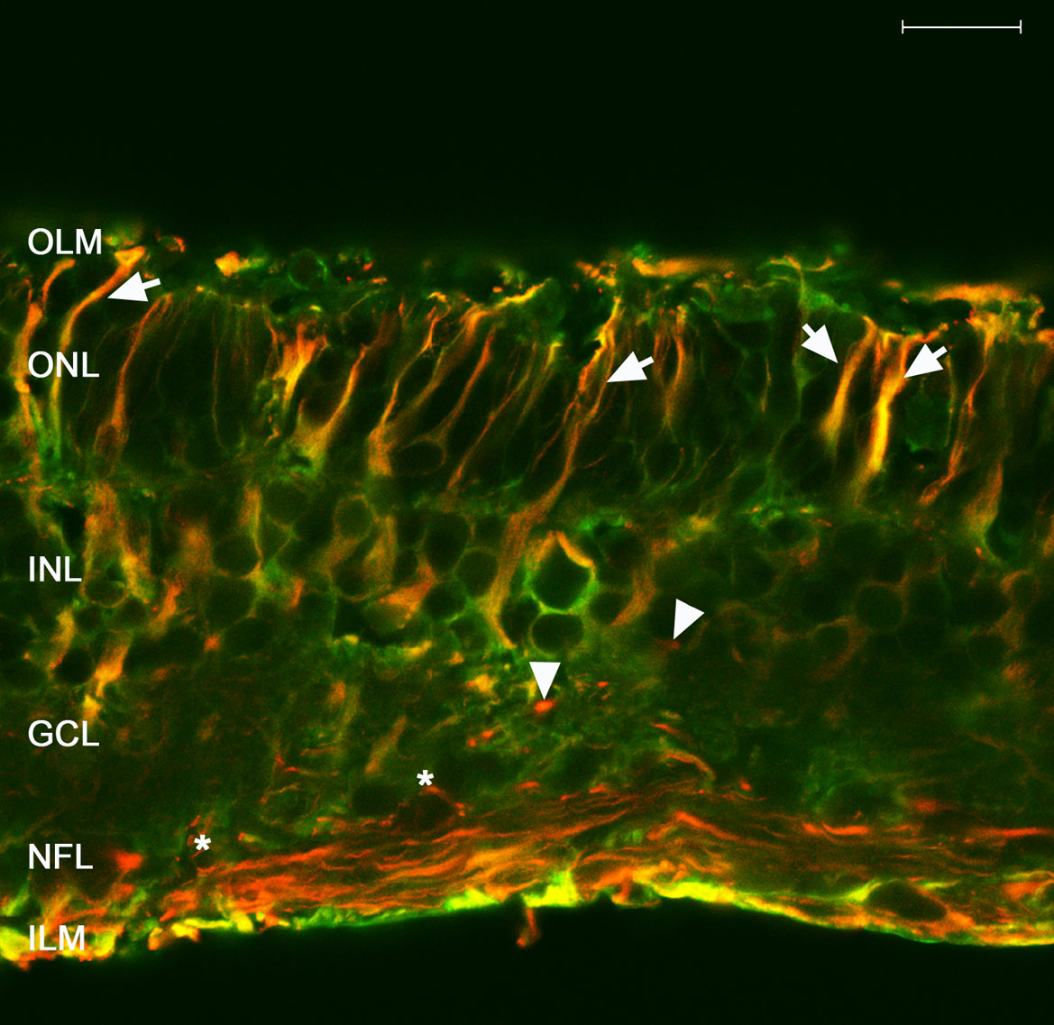
 Figure 6 of
Fernandez-Bueno, Mol Vis 2008; 14:2148-2156.
Figure 6 of
Fernandez-Bueno, Mol Vis 2008; 14:2148-2156.