Figure 5. Immunofluorescence staining of
semithin sections from explants cocultured with the mononuclear
fraction. Antibody against glial fibrillary acidic protein (GFAP; red)
and DAPI dye (blue) were used. At 3 (A), 6 (B), and 9 (C)
days of culture, the Müller cells were wider and more positive for GFAP
than controls. After 9 days in culture (C), numerous Müller cell
GFAP+ processes invaded the subretinal space, distributed in several
fibrous layers (arrows). Scale bar: 20 µm. Abbreviations: GCL is
ganglion cell layer; ILM is inner limiting membrane; INL is inner
nuclear layer; NFL: nerve fiber layer; ONL is outer nuclear layer.
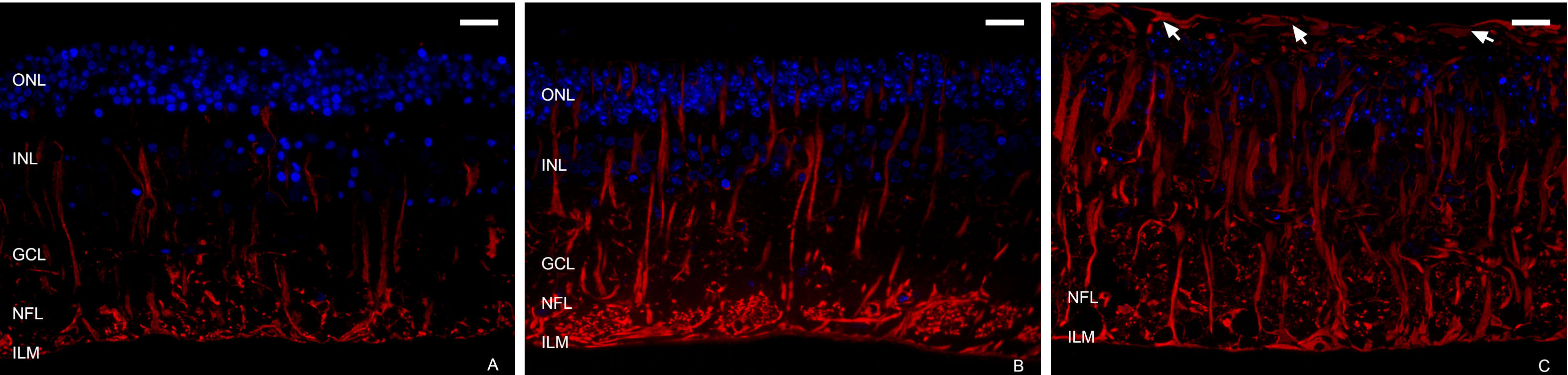
 Figure 5 of Fernandez-Bueno, Mol Vis 2008; 14:2148-2156.