Figure 3. Immunofluorescence staining of cryosections from 6 day control explants. As seen in confocal images of cryostat sections,
antibodies against glial fibrillary acidic protein (GFAP; red) identified glial intermediate filaments (IF), and antibodies
against cellular retinaldehyde-binding protein (CRALBP; green) identified a retinoid-binding protein present in Müller cells.
CRALBP labeling was more concentrated at the neuroretinal outer layers. Colocalization of both antibodies (yellow) marked
Müller cells GFAP+. In control explants, only some Müller cells showed GFAP expression in the cytoplasm that reached the outer
limiting membrane (OLM; arrows). Astrocyte cell bodies and GFAP+ extensions were located along the nerve fiber layer (NFL;
arrowheads). Scale bar equals 20 µm. Abbreviations: ganglion cell layer (GCL); inner limiting membrane (ILM); inner nuclear
layer (INL); outer nuclear layer (ONL).
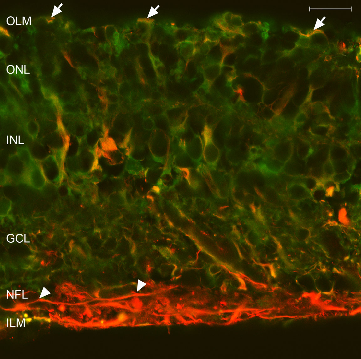
 Figure 3 of
Fernandez-Bueno, Mol Vis 2008; 14:2148-2156.
Figure 3 of
Fernandez-Bueno, Mol Vis 2008; 14:2148-2156.