Figure 2. Immunofluorescence staining of
semithin sections from control explants. Antibodies against glial
fibrillary acidic protein (GFAP; red) were used to identify glial IF.
DAPI dye (blue) was used to label nuclei. In newly detached samples (A),
GFAP was present in the end feet of Müller cells (inner limiting
membrane, ILM) and in astrocytes (nerve fiber layer, NFL). The outer
nuclear layer (ONL), inner nuclear layer (INL), and ganglion cell layer
(GCL) were identified with DAPI dye. At 3 days of culture (B),
GFAP was detectable throughout the Müller cell cytoplasm, from the ILM
to the ONL (arrows). After 6 days of culture (C), the Müller
cells were wider and their GFAP+ processes reached the outer limiting
membrane (OLM; note arrows). After 9 days in culture, in explants that
maintained the retinal structure (D), labeled processes extended
beyond the OLM and began to create a continuous layer in the subretinal
space (arrows). In samples that lost the characteristic retinal
organization (E), nuclei of surviving cells and GFAP+ extensions
were randomly distributed, appearing over the OLM (arrows). Scale bar
equals 20 µm.
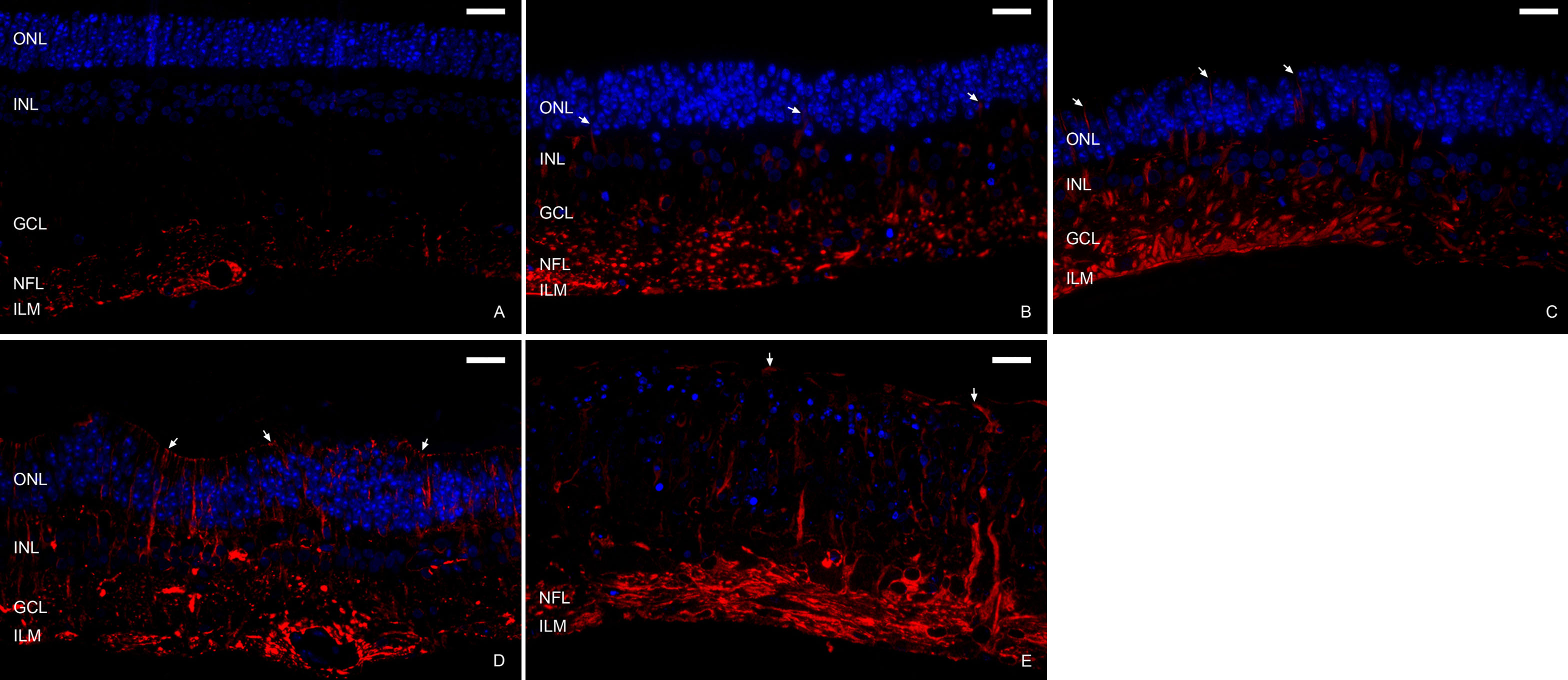
 Figure 2 of Fernandez-Bueno, Mol Vis 2008; 14:2148-2156.