Figure 1. Toluidine blue staining of
semithin sections from control explants. Neuroretinal morphology was
preserved after experimental retinal detachment (A). At day 3 of
culture (B), photoreceptor outer segments (OS) were truncated
and disrupted and the neuroretina began to degenerate and become
thinner. Vacuolization of the ganglion cell layer (GCL) and inner
nuclear layer (INL) was apparent. The outer nuclear layer (ONL) showed
6–7 rows of photoreceptor nuclei as in post-detached specimens, but at
this time point they were loosely distributed. Fragmented OS were
present in the subretinal space (ellipsoid area). After 6 days of
culture (C), the number of rows of nuclei was reduced in the
INL, but remained constant in the ONL. The neuroretinal thickness
continued to decrease, mainly due to narrowing of the plexiform layers.
Fragmented OS remained present in the subretinal space (ellipsoid
area). After 9 days in culture, explants that maintained the retinal
architecture (D) revealed a lower packing density of cells, and
there was a marked reduction in the number of nuclei in the INL. The
plexiform layers almost disappeared and a tissue layer was present
outside the outer limiting membrane (OLM; arrows). In explants that
showed cellular disorganization (E), nuclei were randomly
arranged, appearing outside the OLM and comprised a new multinuclear
tissue layer in the subretinal space (arrows). Comparing (A-E)
images, the retinal thinning during the culture is apparent. On days 3,
6, and 9, the ONL looked progressively thicker (B-D). Scale bar
equals 20 µm. Abbreviations: inner nuclear layer (INL); nerve fiber
layer (NFL).
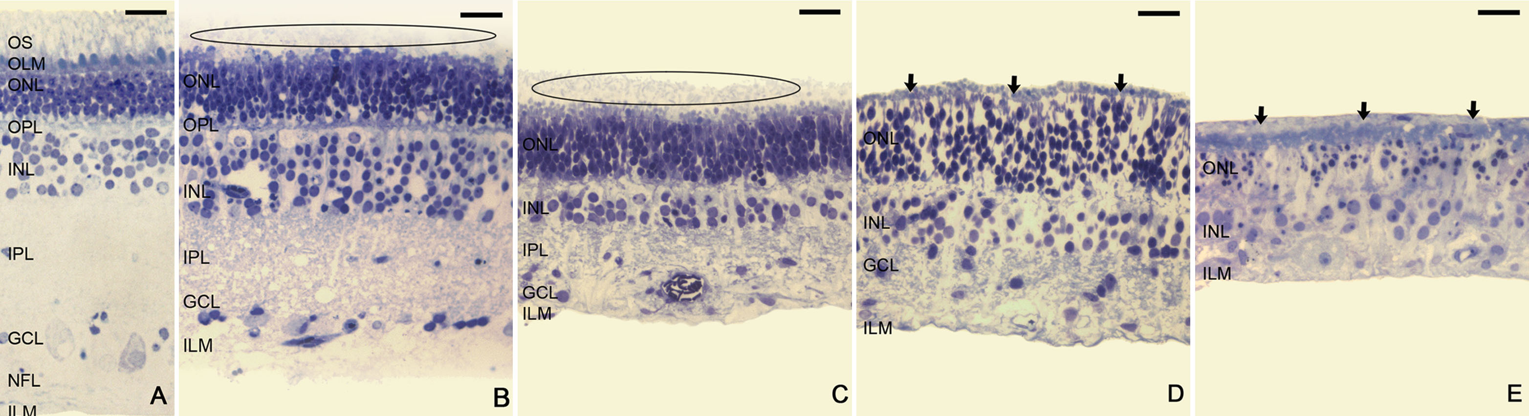
 Figure 1 of Fernandez-Bueno, Mol Vis 2008; 14:2148-2156.