Figure 5. Immunocytochemical analysis of
differentiated cells from spheres derived from the peripheral cornea.
Cells migrating out from the spheres express α-SMA, MAP2, and NSE,
indicating that the colonies contain differentiated mesenchymal and
neuronal cells. There is no staining with the control IgG. Scale
bar=100 μm.
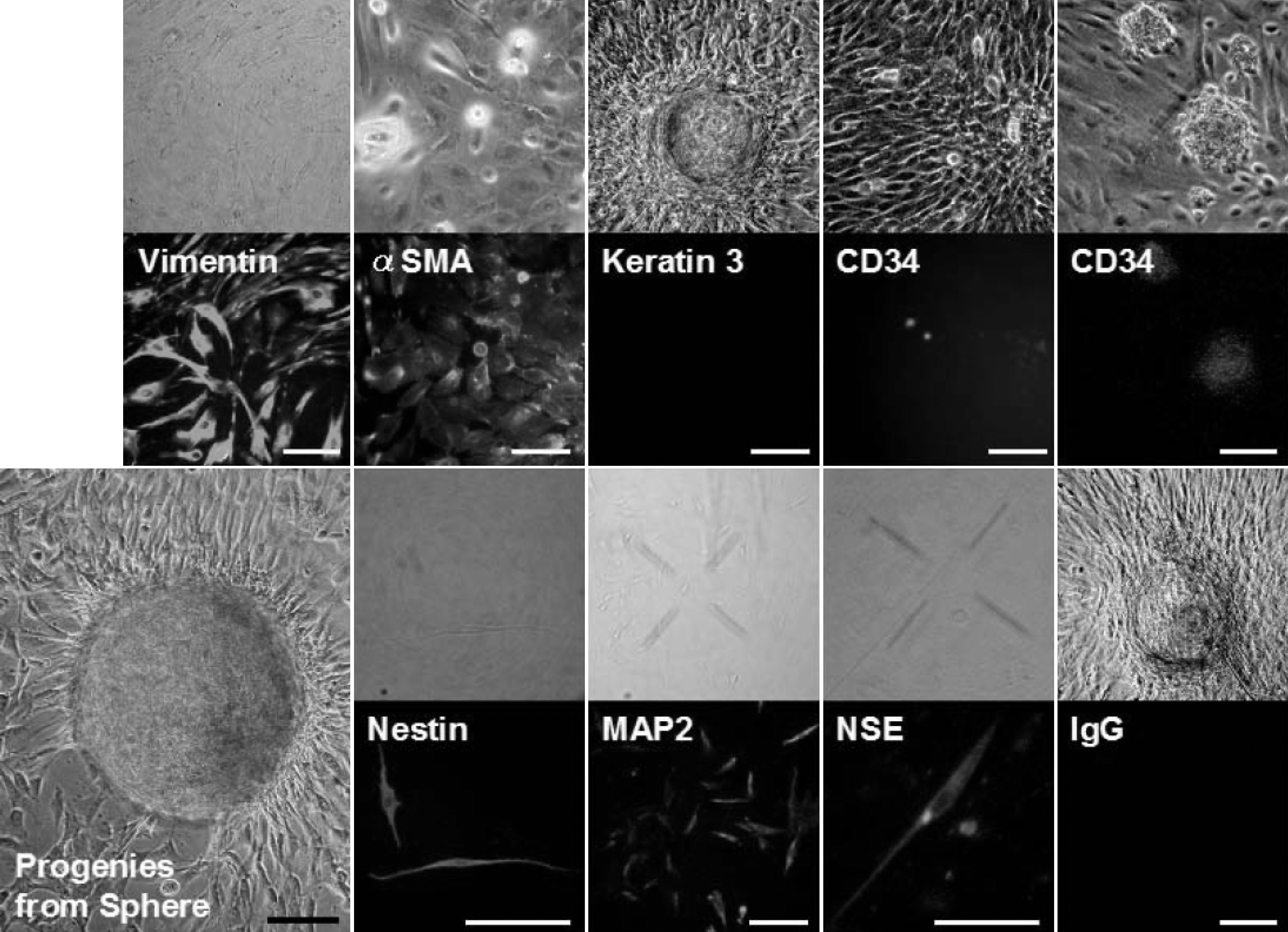
 Figure 5 of Mimura, Mol Vis 2008; 14:197-203.