Figure 4. Immunocytochemical analysis of
sphere colonies from the peripheral stroma on day 7. Bright-field
images and immunostaining of spheres are shown. The spheres were
stained for vimentin (a mesenchymal cell marker), \alpha-smooth muscle
actin (α-SMA, a mesenchymal cell marker), cytokeratin 3 (a
differentiated epithelial cell marker), nestin (a neural stem cell
marker), microtubule-associated protein 2 (MAP2, a differentiated
neural cell marker), neuron-specific enolase (NSE, a differentiated
neural cell marker, and CD34 (a stem cell marker). Each colony is also
labeled by BrdU. As a negative control, IgG was used instead of the
primary antibody. Scale bar=100 μm.
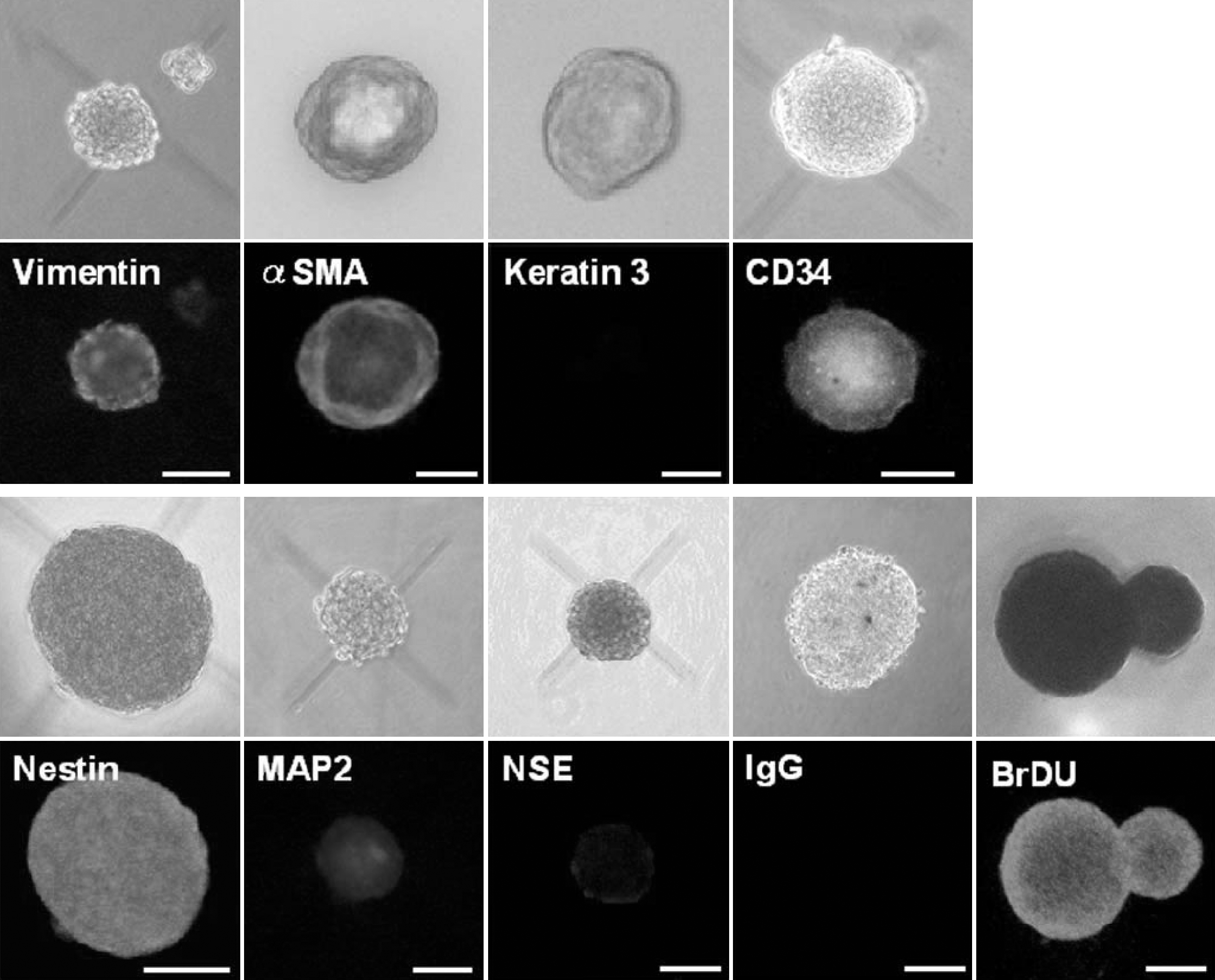
 Figure 4 of Mimura, Mol Vis 2008; 14:197-203.