Figure 3. Formation of secondary sphere. (A,
B) Secondary spheres were generated after the dissociation of
primary spheres derived from peripheral or central keratocytes. Scale
bar=100 μm. (C) The re-plating efficiency from primary to
secondary colonies was higher for spheres derived from the peripheral
stroma than for those from the central stroma (p=0.000025, unpaired t-test).
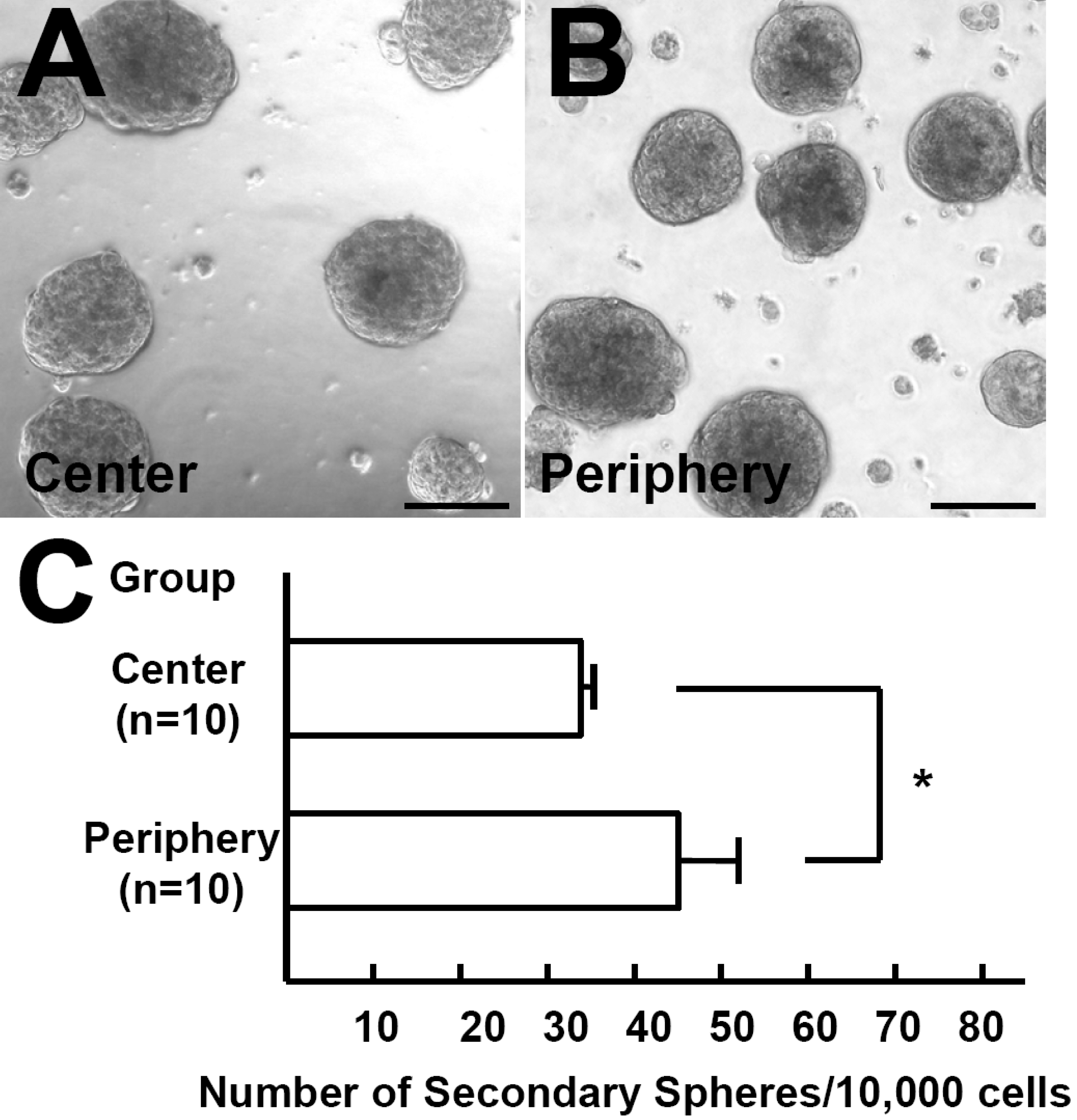
 Figure 3 of Mimura, Mol Vis 2008; 14:197-203.