Figure 2. Primary sphere formation by
keratocytes from the peripheral and central regions of the rabbit
cornea. (A, B) Stromal cells from the peripheral or
central cornea form spheres. Stromal tissue was disaggregated into
single cells, which were plated at a density of 10 viable cells/μl in a
basal medium containing methylcellulose gel matrix to prevent
reaggregation. More than 99% of the cells were single cells on day 0.
Growth of a representative sphere is shown until day 7. Scale bar=200
μm. (C) The number of primary spheres derived from stromal
tissue was compared between the periphery and center of the cornea. The
number of sphere colonies obtained from samples of the peripheral
stroma (n=10) was significantly higher than that for samples of the
central stroma (n=10) after seven days of culture (The asterisk
indicates that p=0.00021 and unpaired t-test was performed). (D)
The size of primary sphere colonies derived from samples of peripheral
(n=10) and central (n=10) corneal stroma was compared. The mean size of
spheres from both regions gradually increased during culture to exceed
250 μm on day 7 (periphery: 258±63 μm versus center: 203±71 μm after
seven days, mean±SD). n.s.=not significant.
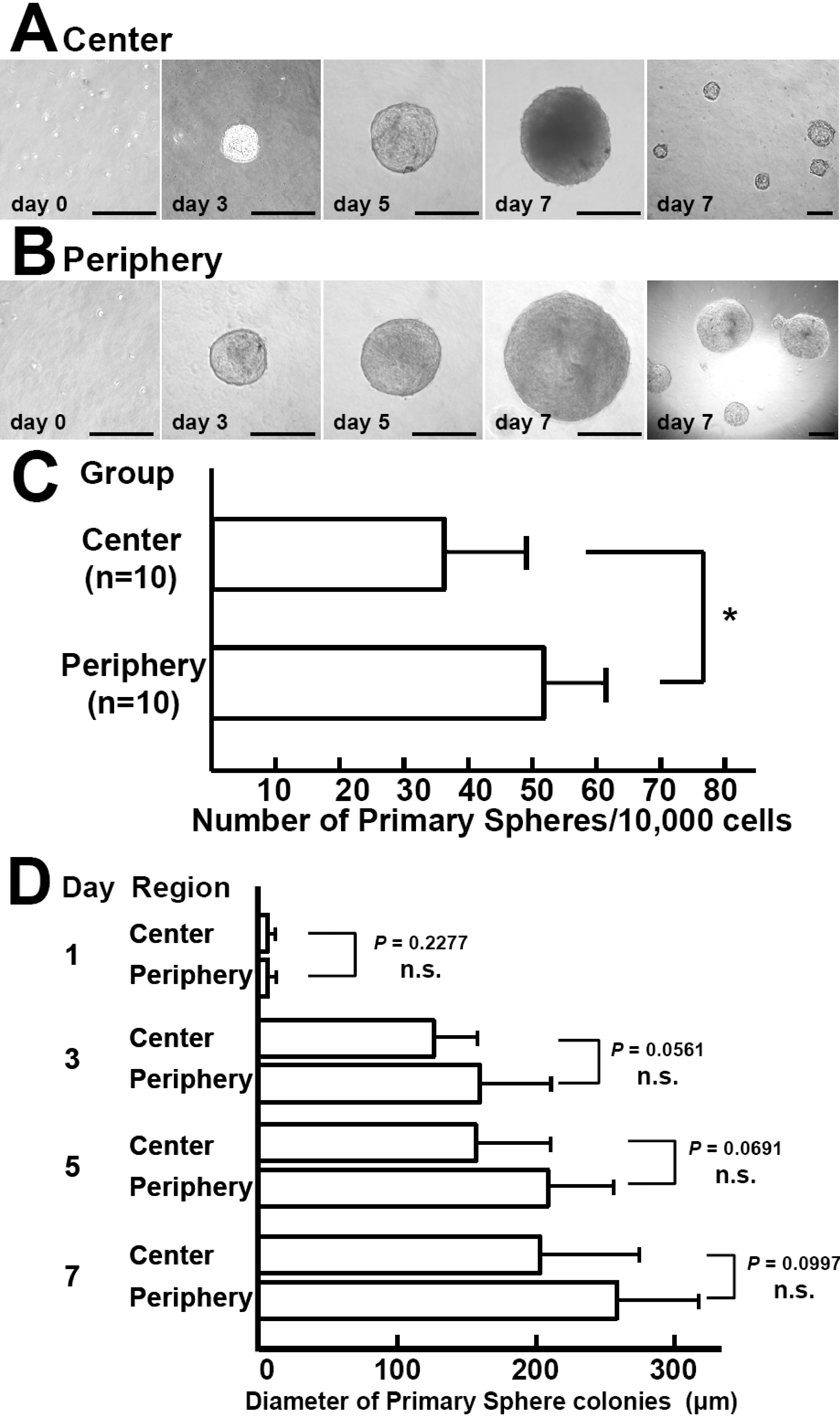
 Figure 2 of Mimura, Mol Vis 2008; 14:197-203.