Figure 1. Anterior view of a rabbit cornea
and a diagram of the corneal epithelium and stroma. The epithelium was
removed from the rabbit corneal stroma by scraping the outer surface of
the cornea while the corneal endothelium and Descemet’s membrane were
peeled away with fine forceps. To compare the distribution and
proliferative capacity of keratocyte precursors obtained from the
central and peripheral regions, stromal keratocytes were isolated from
tissue specimens obtained from both the peripheral (6.0-10.0 mm in
diameter) and central regions (6.0 mm in diameter) using trephines and
forceps.
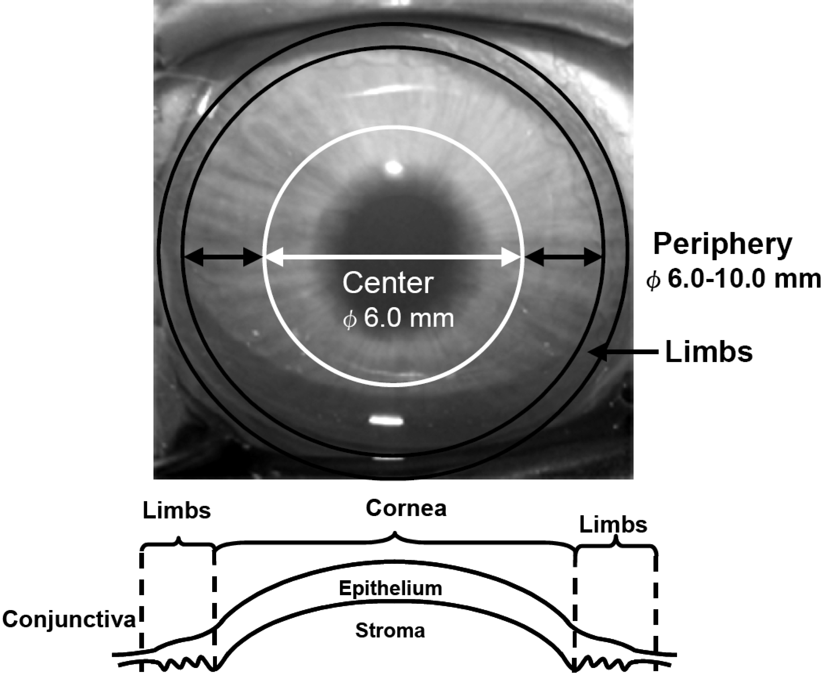
 Figure 1 of Mimura, Mol Vis 2008; 14:197-203.