Figure 7. NGC protein expression in the adult mouse retina. Immunohistochemical analysis was performed with an antiserum raised against
the extracellular part of NGC (red) on retinal sections of 2-month-old wild-type mice (A). Nuclei were stained in blue with DAPI, and images were merged (B). As a negative control, serum from a nonimmunized rabbit was used; the nuclei were stained with DAPI, and the images were
merged (D). C: NGC is predominantly expressed at the apical side of RPE cells. NGC appears in red, nuclei are in blue, and outer segments
of cone photoreceptor cells are in green. Note cone outer segments surrounded by microvilli of RPE cells in yellow (arrows).
This image was obtained by filtering the intensity of the fluorescence down to 6.25% for the red channel, by fixing the one
of the green channel at 100%. Stars denote three nuclei of RPE cells. Abbreviations: retinal pigment epithelium (rpe); outer
nuclear layer (onl); outer plexiform layer (opl); inner nuclear layer (inl); inner plexiform layer (ipl); ganglion cell layer
(gcl). Scale bars equal 30 μm.
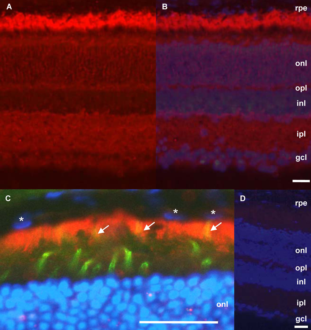
 Figure 7 of
Escher, Mol Vis 2008; 14:2126-2135.
Figure 7 of
Escher, Mol Vis 2008; 14:2126-2135.