Figure 6. NGC protein expression during retinal degeneration in Rpe65−/− mice. A: In the present study, 20 μg of total protein extracts from four pooled retinas of 2-month-old wild-type and Rpe65−/− mice were resolved on a 6% SDS–PAGE and analyzed by western blot. Posttranslational modifications of the NGC full-length
protein resulted in a signal for NGC under appearance of a smear. The asterisk marks the shedded NGC ectodomain of about 75
kDa. B: Total protein extracts were prepared from one retina of 2-, 4-, and 6-months (m)-old wild-type (white bars) and Rpe65-/-mice (black bars). NGC expression was assessed by western blot and subsequently quantified (n=3). The sum of NGC full-length
and ectodomain signal intensities were normalized to α-tubulin expression. NGC expression was statistically different between
wild-type and Rpe65-/- retinas at 2 and 4 months, but not 6 months of age, as assessed by two-way ANOVA (p<0.01) and by Student’s /t/-test.
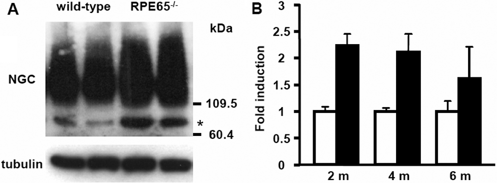
 Figure 6 of
Escher, Mol Vis 2008; 14:2126-2135.
Figure 6 of
Escher, Mol Vis 2008; 14:2126-2135.