Figure 5. YFP expression in differentiated retinal progenitor cells. A: Live images of FIV-YFP infected cells cultured for 14 days under differentiated conditions from a representative experiment.
The cells have adopted a variety of neuronal cell morphologies and have downregulated the expression of YFP (arrows). B: Immunocytochemistry of cells labeled with BrdU (red signal) and cultured under differentiating conditions. BrdU-negative
cells downregulated the expression of YFP (arrows). The nuclei of all cells were stained with DAPI (blue signal). The results
were examined using a Zeiss Axiovert fluorescence microscope, and images from the representative experiments are shown.
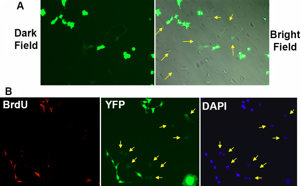
 Figure 5 of
Janic, Mol Vis 2008; 14:2117-2125.
Figure 5 of
Janic, Mol Vis 2008; 14:2117-2125.