Figure 4. Multipotentiality. A: Retinal progenitor cells (RPCs) transduced with feline immunodeficiency virus (FIV) for yellow fluorescent protein (YFP)
were cultured under differentiated conditions for 14 days to give rise to neuronal and glial cell types. Cells that stained
positive for microtubule-associated protein 2 (MAP-2), opsin, and glial fibrillary acidic protein (GFAP) were analyzed by
confocal microscopy, and signaling displayed in red. Green signal was YFP, and blue signal was DAPI staining for nuclei. B: Quantitative analysis of cells positive for MAP-2, opsin, and GFAP in RPC group infected with or without FIV-YFP. At least
100 cells were counted from three independent experiments. Results are expressed as mean±SEM; p>0.05 (Student’s t-test; FIV-YFP
versus control).
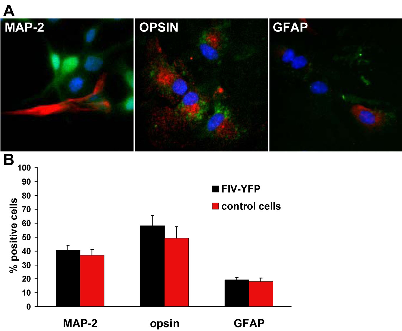
 Figure 4 of
Janic, Mol Vis 2008; 14:2117-2125.
Figure 4 of
Janic, Mol Vis 2008; 14:2117-2125.