Figure 3. Long-term expression of YFP reporter in FIV-transduced RPCs. Dissociated RPCs were transduced with feline immunodeficiency
virus (FIV) expressing yellow fluorescent protein (YFP) and plated at a low density of 10 cells/μl in the complete growth
medium to generate clonal spheres. Cells were monitored, and images captured at days 1, 4, and 7 post-dissociation by the
inverted fluorescence microscope to detect the presence of secondary RPC spheres. Long-term expression of YFP reporter was
still detected after 10 passages. Shown are live-phase contrast cell images (fluorescence and bright field) from the representative
culture.
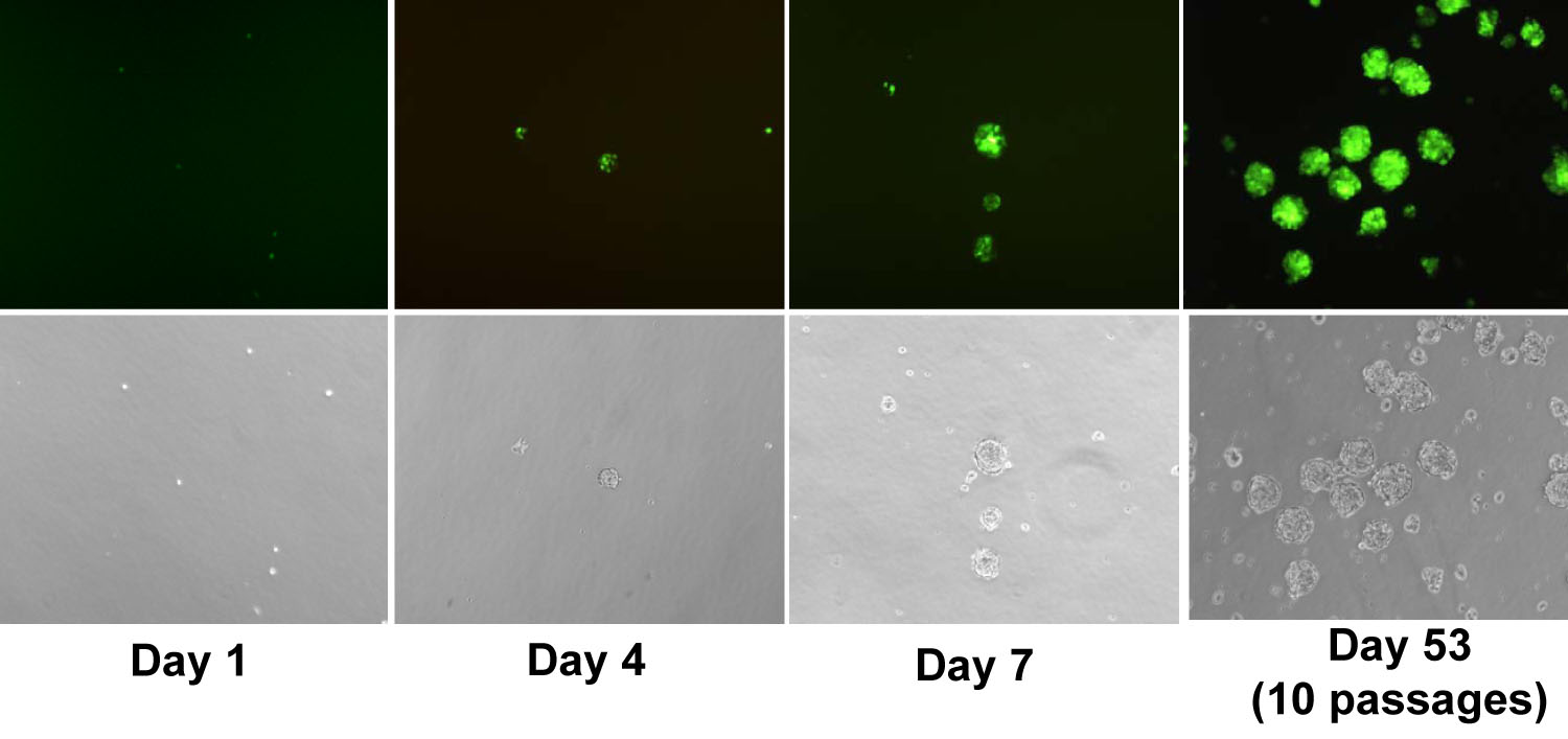
 Figure 3 of
Janic, Mol Vis 2008; 14:2117-2125.
Figure 3 of
Janic, Mol Vis 2008; 14:2117-2125.