Figure 2. Nestin expression in
FIV-transduced retinal progenitor cells. A: Immunocytochemistry
for nestin expression (red signal) in retinal progenitor cell (RPC)
spheres cultured under proliferating conditions. The nuclei of all
cells were stained with DAPI (blue signal) and analyzed using a Zeiss
Axiovert 200 m fluorescence microscope. B: Quantitative
analysis of nestin-positive cells in feline immunodeficiency virus
(FIV) for yellow fluorescent protein (YFP)-infected and noninfected
RPCs. The percentage of nestin-expressing cells was determined by
counting at least 100 cells in three independent experiments. Results
are expressed as mean±SEM; p<0.05 (Student’s t-test; FIV-YFP versus
control).
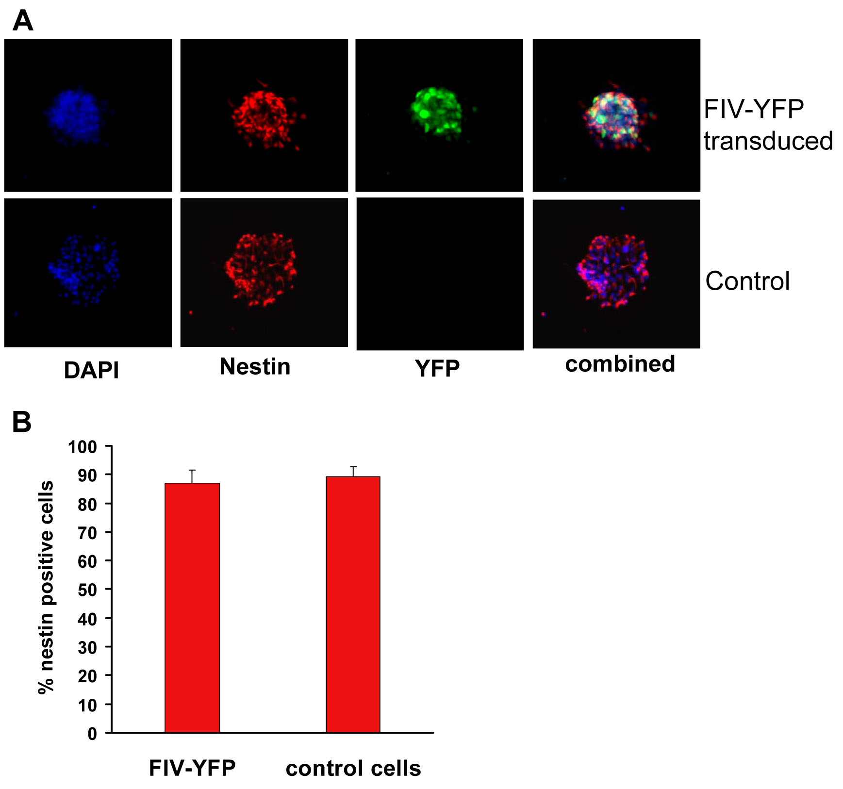
 Figure 2 of Janic, Mol Vis 2008; 14:2117-2125.