Figure 2. Wild-type/mutant myocilin
co-expression significantly increases the amount of extracellular
mutant myocilin compared to single mutant expression. HEK-293T cells
were transiently transfected with 200 ng of cDNA constructs encoding 5
different myocilin-myc mutants or wild-type myocilin-myc (A). To
model the heterozygous expression of myocilin mutants, cells were
co-transfected with 200 ng of cDNAs encoding each mutant myocilin-myc
and 200 ng of a cDNA construct encoding wild-type myocilin-HA.
Twenty-four hours after transfection, the recombinant mutant-myc (C)
and wild-type-HA myocilins (E) in both the culture medium and
cells were analyzed by 10% polyacrylamide SDS-PAGE and western blot
using either an anti-myc or an anti-HA monoclonal antibody,
respectively. Arrows and arrowheads indicate the position of
full-length myocilin (55 kDa), and the 35 kDa processed
olfactomedin-containing fragment, respectively. Molecular weight
markers are shown on the left. B, D and F:
Densitometric quantitation of full-length myocilin and its processed
olfactomedin-containing fragment, detected in A (bars with
oblique lines), C (solid bars), and E (dotted
bars). Red bars represent the percentage of extracellular unprocessed
full-length or truncated Q368X myocilin (extracellular myocilin/total
myocilin), blue bars correspond to the extracellular
olfactomedin-containing fragment (extracellular 35 kDa myocilin/total
myocilin), and yellow bars represent intracellular full-length myocilin
or the truncated Q368X form (intracellular myocilin/total myocilin),
respectively. Total myocilin in each experiment was calculated as the
sum of all the myocilin signals in both the culture medium and the
cellular fraction. Error bars represent the S.E. of triplicate
experiments. To facilitate the comparison of the percentage of
extracellular unprocessed full-length or truncated Q368X myocilin, the
corresponding bars form panels B and D (G) or
panel F (H) are represented together. Numbers above bars
in panel G indicate the fold-increase of full-length myocilin
in cotransfections versus single transfections. Statistical
significance as compared with full-length wild-type myocilin, was
calculated using the Student’s test. The asterisk indicates a
p<0.05, the double asterisk indicates a p<0.01 and the triple
asterisk indicates a p<0.001.
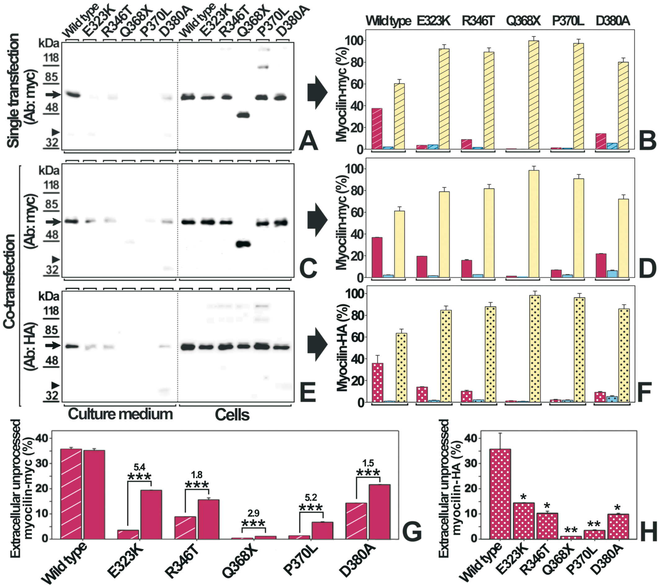
 Figure 2 of Aroca-Aguilar, Mol Vis 2008; 14:2097-2108.