Figure 1. Scheme of wild-type and mutant myocilin cDNA constructs used in this study. The localization of different domains and epitopes
of myocilin is indicated by rectangles filled with different patterns. Numbers correspond to positions in the myocilin amino
acid sequence.
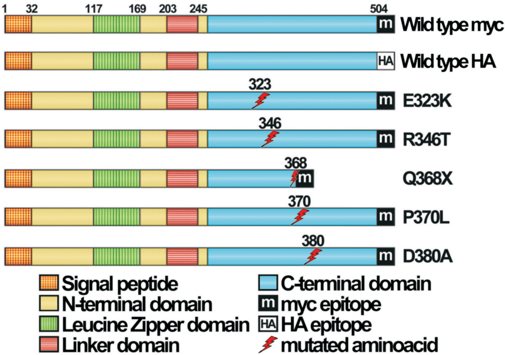
 Figure 1 of
Aroca-Aguilar, Mol Vis 2008; 14:2097-2108.
Figure 1 of
Aroca-Aguilar, Mol Vis 2008; 14:2097-2108.