Figure 5. rAV-GFP transduction of
organ-cultured normal corneas. Upper left, live cornea; other panels,
transverse corneal sections (combined fluorescence and transmitted
light). Mostly epithelial (A,B) and some endothelial
cells (D) are transgene-positive, whereas stromal keratocytes (C)
are negative. e, epithelium; s, stroma; DM, Descemet’s membrane. Direct
fluorescence. Bar=20 μm.
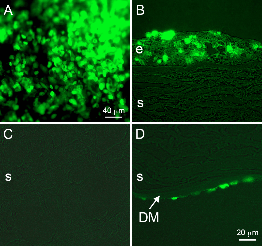
 Figure 5 of Liu, Mol Vis 2008; 14:2087-2096.