Figure 3. rAAV-GFP transduction of rabbit corneas. Upper row, rAAV-GFP5; strong signal is seen in the epithelium (e), stromal cells
(s), and endothelium (en). Epithelial, stromal, and endothelial cells are positive for GFP after rAAV treatment. Lower row,
untreated rabbit corneas as a negative control showing background fluorescence. DM indicates Descemet’s membrane. GFP was
visualized using confocal microscopy and alkaline phosphatase fluorescent detection system. Bar=20 μm.
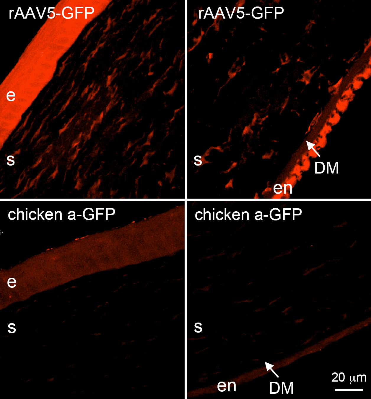
 Figure 3 of
Liu, Mol Vis 2008; 14:2087-2096.
Figure 3 of
Liu, Mol Vis 2008; 14:2087-2096.