Figure 1 of
Liu, Mol Vis 2008; 14:2087-2096.
Figure 1.
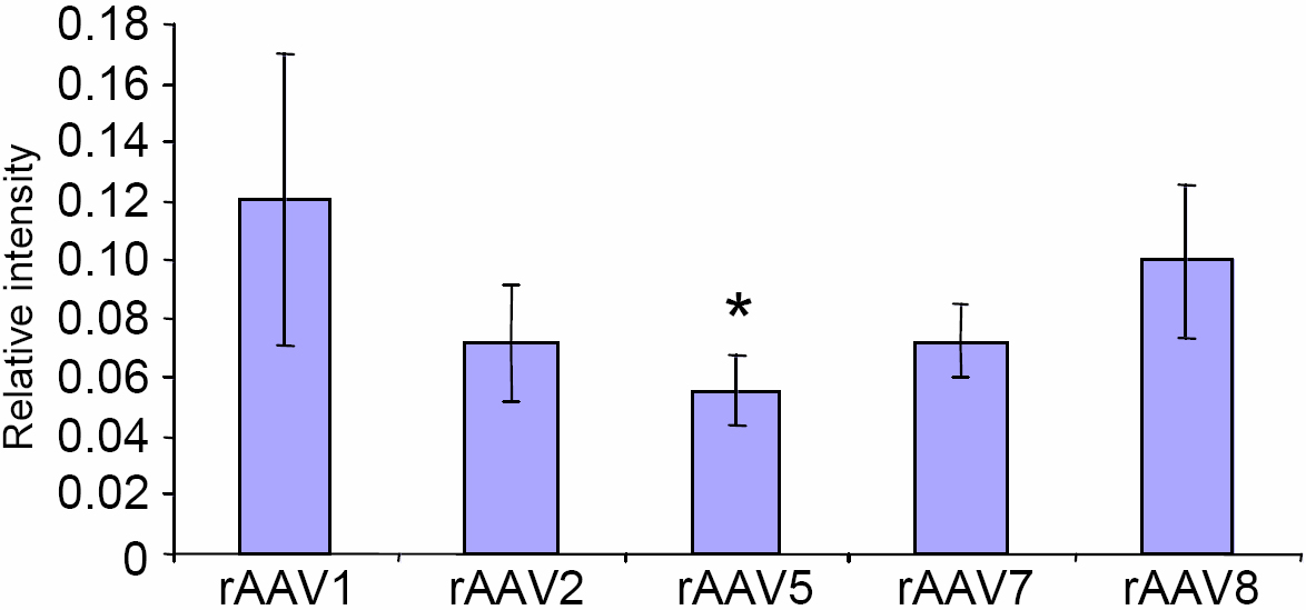
Quantitation of the GFP intensity of staining in corneal epithelium. The strongest signal is observed with rAAV1 and rAAV8, with other serotypes being weaker. The asterisk indicates a p=0.03 (rAAV5 versus rAAV1).
 Figure 1 of
Liu, Mol Vis 2008; 14:2087-2096.
Figure 1 of
Liu, Mol Vis 2008; 14:2087-2096.