Figure 4. Docking models of Prox1-PCNA interaction. A: Ribbon diagram of the complete trimeric PCNA structure (blue) docked to the homeo-Prospero domain of Prox1 (red). Interface
residues are depicted as van der Walls spheres and the PIP box of Prox1 is shown in yellow. B: Ribbon diagram of the interface between the IDCL of PCNA (blue) and Prox1 (red). Residue F693 (green) faces away from the
surface and is inaccessible for interaction with PCNA; whereas residues F692 (yellow), Q686 (red), and Leu 689 (magenta) are
well positioned to directly interact with the IDCL of PCNA.
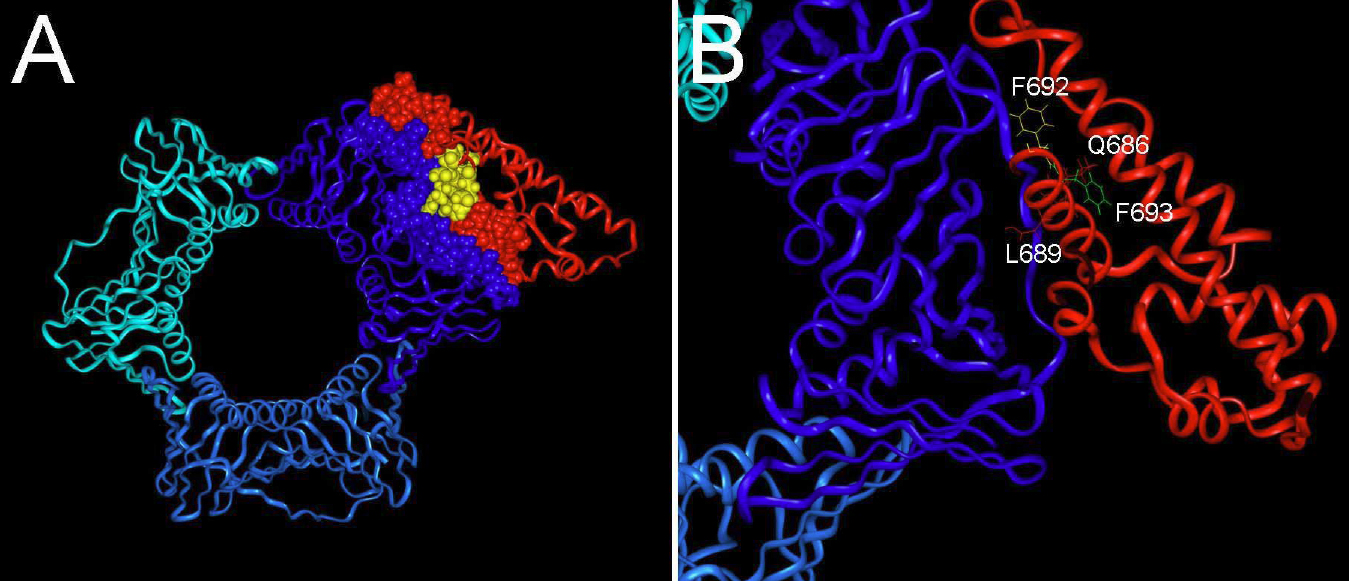
 Figure 4 of
Chen, Mol Vis 2008; 14:2076-2086.
Figure 4 of
Chen, Mol Vis 2008; 14:2076-2086.