Figure 2. Fundus appearance of a patient with Usher syndrome type II. Fundal photographs from a proband (family F5) show typical retinal
degeneration with attenuation of the retinal vessels, irregular pigment clumps in the retina, and waxy pallor of the optic
nerve head.
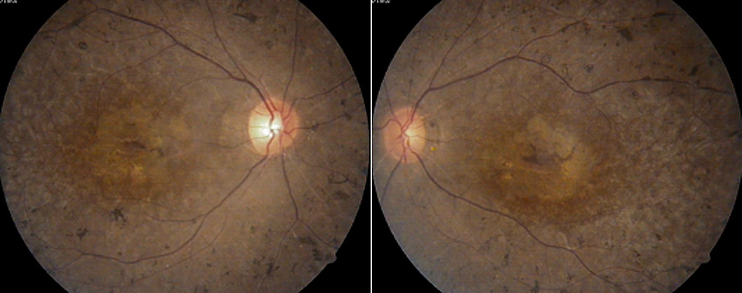
 Figure 2 of
Dai, Mol Vis 2008; 14:2067-2075.
Figure 2 of
Dai, Mol Vis 2008; 14:2067-2075.